{"title":"全膝关节置换术后踝关节和距下关节的冠状面排列是否恢复正常?","authors":"Katsuki Yamaguchi, Tatsuya Sakai, Masanori Fujii, Satoshi Takashima, Shuichi Eto, Yosuke Matsumura, Satomi Nagamine, Hirofumi Tanaka","doi":"10.1186/s43019-025-00272-7","DOIUrl":null,"url":null,"abstract":"<p><strong>Background: </strong>Total knee arthroplasty (TKA) alters the lower extremity alignment, potentially affecting adjacent joints such as the ankle and subtalar joints. However, the relationship between changes in hindfoot alignment and ankle osteoarthritis (OA) after TKA remains incompletely understood. The purpose of this study was to clarify whether ankle and subtalar alignment normalizes after TKA and to identify factors associated with persistent malalignment.</p><p><strong>Methods: </strong>We retrospectively analyzed 331 patients who underwent unilateral mechanical alignment (MA) TKA for knee osteoarthritis. A control group of 40 healthy subjects was used to define normal alignment ranges. Whole-leg anteroposterior weight-bearing radiographs were obtained preoperatively and 2 months postoperatively. Alignment parameters included the hip-knee-ankle angle (HKA), tibiotalar tilt angle (TTA), tibial plafond inclination angle (TPIA), talar inclination angle (TIA), and hindfoot alignment angle (HAA). Pre- and postoperative values were compared using the Wilcoxon signed-rank test, and changes in the proportion of patients within the normal range were determined. Wilcoxon rank-sum tests and chi-squared tests were used for group comparisons, and multivariate logistic regression identified independent predictors of persistent malalignment.</p><p><strong>Results: </strong>HKA improved after TKA (-12° to -2.0°), with corresponding improvements in TPIA (99° to 94°) and TIA (99° to 95°) (all p < 0.001), indicating a significant correction toward neutral alignment. The proportion of patients within normal range increased postoperatively from 16% to 85% for HKA, 26% to 67% for TPIA, 24% to 64% for TIA, and 65% to 73% for HAA. Multivariate analysis identified ankle OA (odds ratio [OR] = 6.62 for TTA), female sex (OR = 2.32 for TPIA; OR = 3.19 for TIA), and varus knee alignment (OR = 2.81 for TIA) as independent predictors of persistent malalignment.</p><p><strong>Conclusions: </strong>MA-TKA facilitates partial normalization of coronal hindfoot alignment, particularly at the tibial plafond and talus. However, female sex, varus knee deformity, and pre-existing ankle OA independently limit full correction. These findings highlight the biomechanical interdependence between the knee and hindfoot and may guide surgical decision-making and patient-specific alignment strategies.</p>","PeriodicalId":36317,"journal":{"name":"Knee Surgery and Related Research","volume":"37 1","pages":"20"},"PeriodicalIF":4.4000,"publicationDate":"2025-05-08","publicationTypes":"Journal Article","fieldsOfStudy":null,"isOpenAccess":false,"openAccessPdf":"https://www.ncbi.nlm.nih.gov/pmc/articles/PMC12063276/pdf/","citationCount":"0","resultStr":"{\"title\":\"Does the coronal plane alignment of the ankle and subtalar joints normalize after total knee arthroplasty?\",\"authors\":\"Katsuki Yamaguchi, Tatsuya Sakai, Masanori Fujii, Satoshi Takashima, Shuichi Eto, Yosuke Matsumura, Satomi Nagamine, Hirofumi Tanaka\",\"doi\":\"10.1186/s43019-025-00272-7\",\"DOIUrl\":null,\"url\":null,\"abstract\":\"<p><strong>Background: </strong>Total knee arthroplasty (TKA) alters the lower extremity alignment, potentially affecting adjacent joints such as the ankle and subtalar joints. However, the relationship between changes in hindfoot alignment and ankle osteoarthritis (OA) after TKA remains incompletely understood. The purpose of this study was to clarify whether ankle and subtalar alignment normalizes after TKA and to identify factors associated with persistent malalignment.</p><p><strong>Methods: </strong>We retrospectively analyzed 331 patients who underwent unilateral mechanical alignment (MA) TKA for knee osteoarthritis. A control group of 40 healthy subjects was used to define normal alignment ranges. Whole-leg anteroposterior weight-bearing radiographs were obtained preoperatively and 2 months postoperatively. Alignment parameters included the hip-knee-ankle angle (HKA), tibiotalar tilt angle (TTA), tibial plafond inclination angle (TPIA), talar inclination angle (TIA), and hindfoot alignment angle (HAA). Pre- and postoperative values were compared using the Wilcoxon signed-rank test, and changes in the proportion of patients within the normal range were determined. Wilcoxon rank-sum tests and chi-squared tests were used for group comparisons, and multivariate logistic regression identified independent predictors of persistent malalignment.</p><p><strong>Results: </strong>HKA improved after TKA (-12° to -2.0°), with corresponding improvements in TPIA (99° to 94°) and TIA (99° to 95°) (all p < 0.001), indicating a significant correction toward neutral alignment. The proportion of patients within normal range increased postoperatively from 16% to 85% for HKA, 26% to 67% for TPIA, 24% to 64% for TIA, and 65% to 73% for HAA. Multivariate analysis identified ankle OA (odds ratio [OR] = 6.62 for TTA), female sex (OR = 2.32 for TPIA; OR = 3.19 for TIA), and varus knee alignment (OR = 2.81 for TIA) as independent predictors of persistent malalignment.</p><p><strong>Conclusions: </strong>MA-TKA facilitates partial normalization of coronal hindfoot alignment, particularly at the tibial plafond and talus. However, female sex, varus knee deformity, and pre-existing ankle OA independently limit full correction. These findings highlight the biomechanical interdependence between the knee and hindfoot and may guide surgical decision-making and patient-specific alignment strategies.</p>\",\"PeriodicalId\":36317,\"journal\":{\"name\":\"Knee Surgery and Related Research\",\"volume\":\"37 1\",\"pages\":\"20\"},\"PeriodicalIF\":4.4000,\"publicationDate\":\"2025-05-08\",\"publicationTypes\":\"Journal Article\",\"fieldsOfStudy\":null,\"isOpenAccess\":false,\"openAccessPdf\":\"https://www.ncbi.nlm.nih.gov/pmc/articles/PMC12063276/pdf/\",\"citationCount\":\"0\",\"resultStr\":null,\"platform\":\"Semanticscholar\",\"paperid\":null,\"PeriodicalName\":\"Knee Surgery and Related Research\",\"FirstCategoryId\":\"1085\",\"ListUrlMain\":\"https://doi.org/10.1186/s43019-025-00272-7\",\"RegionNum\":0,\"RegionCategory\":null,\"ArticlePicture\":[],\"TitleCN\":null,\"AbstractTextCN\":null,\"PMCID\":null,\"EPubDate\":\"\",\"PubModel\":\"\",\"JCR\":\"Q2\",\"JCRName\":\"Medicine\",\"Score\":null,\"Total\":0}","platform":"Semanticscholar","paperid":null,"PeriodicalName":"Knee Surgery and Related Research","FirstCategoryId":"1085","ListUrlMain":"https://doi.org/10.1186/s43019-025-00272-7","RegionNum":0,"RegionCategory":null,"ArticlePicture":[],"TitleCN":null,"AbstractTextCN":null,"PMCID":null,"EPubDate":"","PubModel":"","JCR":"Q2","JCRName":"Medicine","Score":null,"Total":0}
Does the coronal plane alignment of the ankle and subtalar joints normalize after total knee arthroplasty?
Background: Total knee arthroplasty (TKA) alters the lower extremity alignment, potentially affecting adjacent joints such as the ankle and subtalar joints. However, the relationship between changes in hindfoot alignment and ankle osteoarthritis (OA) after TKA remains incompletely understood. The purpose of this study was to clarify whether ankle and subtalar alignment normalizes after TKA and to identify factors associated with persistent malalignment.
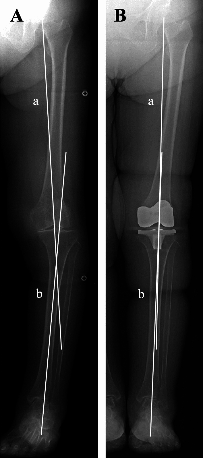
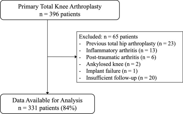
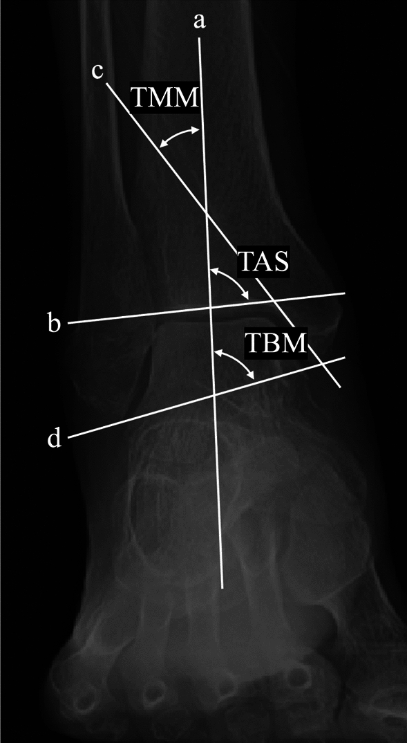
Methods: We retrospectively analyzed 331 patients who underwent unilateral mechanical alignment (MA) TKA for knee osteoarthritis. A control group of 40 healthy subjects was used to define normal alignment ranges. Whole-leg anteroposterior weight-bearing radiographs were obtained preoperatively and 2 months postoperatively. Alignment parameters included the hip-knee-ankle angle (HKA), tibiotalar tilt angle (TTA), tibial plafond inclination angle (TPIA), talar inclination angle (TIA), and hindfoot alignment angle (HAA). Pre- and postoperative values were compared using the Wilcoxon signed-rank test, and changes in the proportion of patients within the normal range were determined. Wilcoxon rank-sum tests and chi-squared tests were used for group comparisons, and multivariate logistic regression identified independent predictors of persistent malalignment.
Results: HKA improved after TKA (-12° to -2.0°), with corresponding improvements in TPIA (99° to 94°) and TIA (99° to 95°) (all p < 0.001), indicating a significant correction toward neutral alignment. The proportion of patients within normal range increased postoperatively from 16% to 85% for HKA, 26% to 67% for TPIA, 24% to 64% for TIA, and 65% to 73% for HAA. Multivariate analysis identified ankle OA (odds ratio [OR] = 6.62 for TTA), female sex (OR = 2.32 for TPIA; OR = 3.19 for TIA), and varus knee alignment (OR = 2.81 for TIA) as independent predictors of persistent malalignment.
Conclusions: MA-TKA facilitates partial normalization of coronal hindfoot alignment, particularly at the tibial plafond and talus. However, female sex, varus knee deformity, and pre-existing ankle OA independently limit full correction. These findings highlight the biomechanical interdependence between the knee and hindfoot and may guide surgical decision-making and patient-specific alignment strategies.

 求助内容:
求助内容: 应助结果提醒方式:


