Matthew J. Valente , Hannah Streett , Randi Turner , Celia O'Brien , Valsin Fournet , Andrew Jansen , Jitender P. Dubey , Benjamin M. Rosenthal , Mark Jenkins , Asis Khan
{"title":"卵囊形态和自身荧光鉴定区分活的和死的球虫寄生虫。","authors":"Matthew J. Valente , Hannah Streett , Randi Turner , Celia O'Brien , Valsin Fournet , Andrew Jansen , Jitender P. Dubey , Benjamin M. Rosenthal , Mark Jenkins , Asis Khan","doi":"10.1016/j.ijpara.2025.04.003","DOIUrl":null,"url":null,"abstract":"<div><div>To assess and mitigate foodborne risk, regulatory agencies and produce growers require the means not merely to detect but moreover determine the viability of foodborne eukaryotic pathogens such as <em>Cyclospora cayetanensis.</em> Viability assessment would also aid those employing live attenuated vaccines against coccidiosis, a major problem in poultry production. Therefore, we sought to identify morphological changes differentiating viable from non-viable coccidian oocysts, employing <em>Eimeria acervulina</em> as a tractable model, enabling empirical validation by means of in vivo challenge infections in the natural chicken host. High resolution microscopic examinations identified granular structures that autofluoresce under UV exposure in dead oocysts, greatly intensifying overall autofluorescence in dead oocysts. We harnessed this intensification as a basis to sort live from dead oocysts using a Fluorescence-Activated Cell Sorting (FACS) cell sorter, validating their distinction by documenting infectivity in chickens using the former, and minimal shedding with the latter. Our rapid, sensitive, and robust assay holds promise for application to other species of coccidia, including those important to livestock and public health.</div></div>","PeriodicalId":13725,"journal":{"name":"International journal for parasitology","volume":"55 8","pages":"Pages 475-484"},"PeriodicalIF":3.2000,"publicationDate":"2025-07-01","publicationTypes":"Journal Article","fieldsOfStudy":null,"isOpenAccess":false,"openAccessPdf":"","citationCount":"0","resultStr":"{\"title\":\"Morphological and autofluorescence assessment of oocysts differentiate live from dead coccidian parasites\",\"authors\":\"Matthew J. Valente , Hannah Streett , Randi Turner , Celia O'Brien , Valsin Fournet , Andrew Jansen , Jitender P. Dubey , Benjamin M. Rosenthal , Mark Jenkins , Asis Khan\",\"doi\":\"10.1016/j.ijpara.2025.04.003\",\"DOIUrl\":null,\"url\":null,\"abstract\":\"<div><div>To assess and mitigate foodborne risk, regulatory agencies and produce growers require the means not merely to detect but moreover determine the viability of foodborne eukaryotic pathogens such as <em>Cyclospora cayetanensis.</em> Viability assessment would also aid those employing live attenuated vaccines against coccidiosis, a major problem in poultry production. Therefore, we sought to identify morphological changes differentiating viable from non-viable coccidian oocysts, employing <em>Eimeria acervulina</em> as a tractable model, enabling empirical validation by means of in vivo challenge infections in the natural chicken host. High resolution microscopic examinations identified granular structures that autofluoresce under UV exposure in dead oocysts, greatly intensifying overall autofluorescence in dead oocysts. We harnessed this intensification as a basis to sort live from dead oocysts using a Fluorescence-Activated Cell Sorting (FACS) cell sorter, validating their distinction by documenting infectivity in chickens using the former, and minimal shedding with the latter. Our rapid, sensitive, and robust assay holds promise for application to other species of coccidia, including those important to livestock and public health.</div></div>\",\"PeriodicalId\":13725,\"journal\":{\"name\":\"International journal for parasitology\",\"volume\":\"55 8\",\"pages\":\"Pages 475-484\"},\"PeriodicalIF\":3.2000,\"publicationDate\":\"2025-07-01\",\"publicationTypes\":\"Journal Article\",\"fieldsOfStudy\":null,\"isOpenAccess\":false,\"openAccessPdf\":\"\",\"citationCount\":\"0\",\"resultStr\":null,\"platform\":\"Semanticscholar\",\"paperid\":null,\"PeriodicalName\":\"International journal for parasitology\",\"FirstCategoryId\":\"3\",\"ListUrlMain\":\"https://www.sciencedirect.com/science/article/pii/S0020751925000657\",\"RegionNum\":2,\"RegionCategory\":\"医学\",\"ArticlePicture\":[],\"TitleCN\":null,\"AbstractTextCN\":null,\"PMCID\":null,\"EPubDate\":\"\",\"PubModel\":\"\",\"JCR\":\"Q1\",\"JCRName\":\"PARASITOLOGY\",\"Score\":null,\"Total\":0}","platform":"Semanticscholar","paperid":null,"PeriodicalName":"International journal for parasitology","FirstCategoryId":"3","ListUrlMain":"https://www.sciencedirect.com/science/article/pii/S0020751925000657","RegionNum":2,"RegionCategory":"医学","ArticlePicture":[],"TitleCN":null,"AbstractTextCN":null,"PMCID":null,"EPubDate":"","PubModel":"","JCR":"Q1","JCRName":"PARASITOLOGY","Score":null,"Total":0}
Morphological and autofluorescence assessment of oocysts differentiate live from dead coccidian parasites
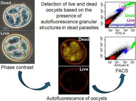
To assess and mitigate foodborne risk, regulatory agencies and produce growers require the means not merely to detect but moreover determine the viability of foodborne eukaryotic pathogens such as Cyclospora cayetanensis. Viability assessment would also aid those employing live attenuated vaccines against coccidiosis, a major problem in poultry production. Therefore, we sought to identify morphological changes differentiating viable from non-viable coccidian oocysts, employing Eimeria acervulina as a tractable model, enabling empirical validation by means of in vivo challenge infections in the natural chicken host. High resolution microscopic examinations identified granular structures that autofluoresce under UV exposure in dead oocysts, greatly intensifying overall autofluorescence in dead oocysts. We harnessed this intensification as a basis to sort live from dead oocysts using a Fluorescence-Activated Cell Sorting (FACS) cell sorter, validating their distinction by documenting infectivity in chickens using the former, and minimal shedding with the latter. Our rapid, sensitive, and robust assay holds promise for application to other species of coccidia, including those important to livestock and public health.
期刊介绍:
International Journal for Parasitology offers authors the option to sponsor nonsubscriber access to their articles on Elsevier electronic publishing platforms. For more information please view our Sponsored Articles page. The International Journal for Parasitology publishes the results of original research in all aspects of basic and applied parasitology, including all the fields covered by its Specialist Editors, and ranging from parasites and host-parasite relationships of intrinsic biological interest to those of social and economic importance in human and veterinary medicine and agriculture.