Mar Fernandez Salamanca, Rita Simões, Malgorzata Deręgowska-Cylke, Pim J van Leeuwen, Henk G van der Poel, Elise Bekers, Marcos A S Guimaraes, Uulke A van der Heide, Ivo G Schoots
{"title":"超越Gleason分级:MRI放射组学区分前列腺癌男性筛状生长和非筛状生长。","authors":"Mar Fernandez Salamanca, Rita Simões, Malgorzata Deręgowska-Cylke, Pim J van Leeuwen, Henk G van der Poel, Elise Bekers, Marcos A S Guimaraes, Uulke A van der Heide, Ivo G Schoots","doi":"10.1007/s10334-025-01251-5","DOIUrl":null,"url":null,"abstract":"<p><strong>Objective: </strong>To differentiate cribriform (GP4Crib+) from non-cribriform growth and Gleason 3 patterns (GP4Crib-/GP3) using MRI.</p><p><strong>Methods: </strong>Two hundred and ninety-one operated prostate cancer men with pre-treatment MRI and whole-mount prostate histology were retrospectively included. T2-weighted, apparent diffusion coefficient (ADC) and fractional blood volume maps from 1.5/3T MRI systems were used. 592 histological GP3, GP4Crib- and GP4Crib+ regions were segmented on whole-mount specimens and manually co-registered to MRI sequences/maps. Radiomics features were extracted, and an erosion process was applied to minimize the impact of delineation uncertainties. A logistic regression model was developed to differentiate GP4Crib+ from GP3/GP4Crib- in the 465 remaining regions. The differences in balanced accuracy between the model and baseline (where all regions are labeled as GP3/GP4Crib-) and 95% confidence intervals (CI) for all metrics were assessed using bootstrapping.</p><p><strong>Results: </strong>The logistic regression model, using the 90th percentile ADC feature with a negative coefficient, showed a balanced accuracy of 0.65 (95% CI: 0.48-0.79), receiver operating characteristic area under the curve (AUC) of 0.75 (95% CI: 0.54-0.92), a precision-recall AUC of 0.35 (95% CI: 0.14-0.68).</p><p><strong>Conclusion: </strong>The radiomics MRI-based model, trained on Gleason sub-patterns segmented on whole-mount specimen, was able to differentiate GP4Crib+ from GP3/GP4Crib- patterns with moderate accuracy. The most dominant feature was the 90th percentile ADC. This exploratory study highlights 90th percentile ADC as a potential biomarker for cribriform growth differentiation, providing insights into future MRI-based risk assessment strategies.</p>","PeriodicalId":18067,"journal":{"name":"Magnetic Resonance Materials in Physics, Biology and Medicine","volume":" ","pages":"817-827"},"PeriodicalIF":2.5000,"publicationDate":"2025-10-01","publicationTypes":"Journal Article","fieldsOfStudy":null,"isOpenAccess":false,"openAccessPdf":"https://www.ncbi.nlm.nih.gov/pmc/articles/PMC12497665/pdf/","citationCount":"0","resultStr":"{\"title\":\"Beyond Gleason grading: MRI radiomics to differentiate cribriform growth from non-cribriform growth in prostate cancer men.\",\"authors\":\"Mar Fernandez Salamanca, Rita Simões, Malgorzata Deręgowska-Cylke, Pim J van Leeuwen, Henk G van der Poel, Elise Bekers, Marcos A S Guimaraes, Uulke A van der Heide, Ivo G Schoots\",\"doi\":\"10.1007/s10334-025-01251-5\",\"DOIUrl\":null,\"url\":null,\"abstract\":\"<p><strong>Objective: </strong>To differentiate cribriform (GP4Crib+) from non-cribriform growth and Gleason 3 patterns (GP4Crib-/GP3) using MRI.</p><p><strong>Methods: </strong>Two hundred and ninety-one operated prostate cancer men with pre-treatment MRI and whole-mount prostate histology were retrospectively included. T2-weighted, apparent diffusion coefficient (ADC) and fractional blood volume maps from 1.5/3T MRI systems were used. 592 histological GP3, GP4Crib- and GP4Crib+ regions were segmented on whole-mount specimens and manually co-registered to MRI sequences/maps. Radiomics features were extracted, and an erosion process was applied to minimize the impact of delineation uncertainties. A logistic regression model was developed to differentiate GP4Crib+ from GP3/GP4Crib- in the 465 remaining regions. The differences in balanced accuracy between the model and baseline (where all regions are labeled as GP3/GP4Crib-) and 95% confidence intervals (CI) for all metrics were assessed using bootstrapping.</p><p><strong>Results: </strong>The logistic regression model, using the 90th percentile ADC feature with a negative coefficient, showed a balanced accuracy of 0.65 (95% CI: 0.48-0.79), receiver operating characteristic area under the curve (AUC) of 0.75 (95% CI: 0.54-0.92), a precision-recall AUC of 0.35 (95% CI: 0.14-0.68).</p><p><strong>Conclusion: </strong>The radiomics MRI-based model, trained on Gleason sub-patterns segmented on whole-mount specimen, was able to differentiate GP4Crib+ from GP3/GP4Crib- patterns with moderate accuracy. The most dominant feature was the 90th percentile ADC. This exploratory study highlights 90th percentile ADC as a potential biomarker for cribriform growth differentiation, providing insights into future MRI-based risk assessment strategies.</p>\",\"PeriodicalId\":18067,\"journal\":{\"name\":\"Magnetic Resonance Materials in Physics, Biology and Medicine\",\"volume\":\" \",\"pages\":\"817-827\"},\"PeriodicalIF\":2.5000,\"publicationDate\":\"2025-10-01\",\"publicationTypes\":\"Journal Article\",\"fieldsOfStudy\":null,\"isOpenAccess\":false,\"openAccessPdf\":\"https://www.ncbi.nlm.nih.gov/pmc/articles/PMC12497665/pdf/\",\"citationCount\":\"0\",\"resultStr\":null,\"platform\":\"Semanticscholar\",\"paperid\":null,\"PeriodicalName\":\"Magnetic Resonance Materials in Physics, Biology and Medicine\",\"FirstCategoryId\":\"3\",\"ListUrlMain\":\"https://doi.org/10.1007/s10334-025-01251-5\",\"RegionNum\":4,\"RegionCategory\":\"医学\",\"ArticlePicture\":[],\"TitleCN\":null,\"AbstractTextCN\":null,\"PMCID\":null,\"EPubDate\":\"2025/4/29 0:00:00\",\"PubModel\":\"Epub\",\"JCR\":\"Q3\",\"JCRName\":\"RADIOLOGY, NUCLEAR MEDICINE & MEDICAL IMAGING\",\"Score\":null,\"Total\":0}","platform":"Semanticscholar","paperid":null,"PeriodicalName":"Magnetic Resonance Materials in Physics, Biology and Medicine","FirstCategoryId":"3","ListUrlMain":"https://doi.org/10.1007/s10334-025-01251-5","RegionNum":4,"RegionCategory":"医学","ArticlePicture":[],"TitleCN":null,"AbstractTextCN":null,"PMCID":null,"EPubDate":"2025/4/29 0:00:00","PubModel":"Epub","JCR":"Q3","JCRName":"RADIOLOGY, NUCLEAR MEDICINE & MEDICAL IMAGING","Score":null,"Total":0}
Beyond Gleason grading: MRI radiomics to differentiate cribriform growth from non-cribriform growth in prostate cancer men.
Objective: To differentiate cribriform (GP4Crib+) from non-cribriform growth and Gleason 3 patterns (GP4Crib-/GP3) using MRI.
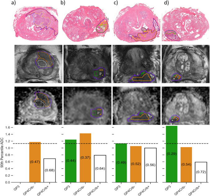
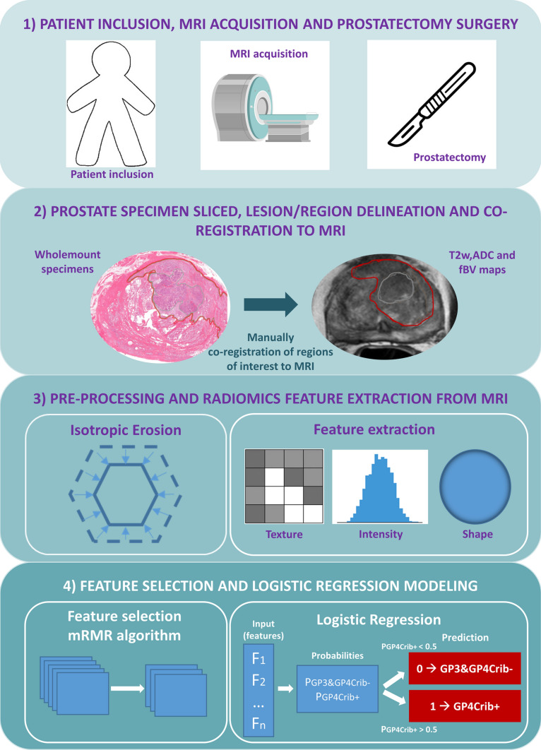
Methods: Two hundred and ninety-one operated prostate cancer men with pre-treatment MRI and whole-mount prostate histology were retrospectively included. T2-weighted, apparent diffusion coefficient (ADC) and fractional blood volume maps from 1.5/3T MRI systems were used. 592 histological GP3, GP4Crib- and GP4Crib+ regions were segmented on whole-mount specimens and manually co-registered to MRI sequences/maps. Radiomics features were extracted, and an erosion process was applied to minimize the impact of delineation uncertainties. A logistic regression model was developed to differentiate GP4Crib+ from GP3/GP4Crib- in the 465 remaining regions. The differences in balanced accuracy between the model and baseline (where all regions are labeled as GP3/GP4Crib-) and 95% confidence intervals (CI) for all metrics were assessed using bootstrapping.
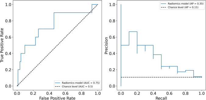
Results: The logistic regression model, using the 90th percentile ADC feature with a negative coefficient, showed a balanced accuracy of 0.65 (95% CI: 0.48-0.79), receiver operating characteristic area under the curve (AUC) of 0.75 (95% CI: 0.54-0.92), a precision-recall AUC of 0.35 (95% CI: 0.14-0.68).
Conclusion: The radiomics MRI-based model, trained on Gleason sub-patterns segmented on whole-mount specimen, was able to differentiate GP4Crib+ from GP3/GP4Crib- patterns with moderate accuracy. The most dominant feature was the 90th percentile ADC. This exploratory study highlights 90th percentile ADC as a potential biomarker for cribriform growth differentiation, providing insights into future MRI-based risk assessment strategies.
期刊介绍:
MAGMA is a multidisciplinary international journal devoted to the publication of articles on all aspects of magnetic resonance techniques and their applications in medicine and biology. MAGMA currently publishes research papers, reviews, letters to the editor, and commentaries, six times a year. The subject areas covered by MAGMA include:
advances in materials, hardware and software in magnetic resonance technology,
new developments and results in research and practical applications of magnetic resonance imaging and spectroscopy related to biology and medicine,
study of animal models and intact cells using magnetic resonance,
reports of clinical trials on humans and clinical validation of magnetic resonance protocols.

 求助内容:
求助内容: 应助结果提醒方式:


