{"title":"深度学习重建胰腺弥散加权成像(DWI)图像质量的改善:与呼吸门控常规DWI的比较","authors":"Kazuki Oyama, Fumihito Ichinohe, Yasuo Adachi, Yoshihiro Kito, Katsuya Maruyama, Minoru Mitsuda, Thomas Benkert, Omar Darwish, Yasunari Fujinaga","doi":"10.1007/s11604-025-01790-w","DOIUrl":null,"url":null,"abstract":"<p><strong>Purpose: </strong>This study aimed to evaluate the efficacy of deep learning-based reconstruction (DLR) in improving pancreatic diffusion-weighted imaging (DWI) quality.</p><p><strong>Materials and methods: </strong>In total, 117 patients (mean age of 68.0 ± 12.9 years) suspected of pancreatic diseases underwent magnetic resonance imaging (MRI) between July and December 2023. MRI sequences included respiratory-gated conventional diffusion-weighted images (RGC-DWIs), respiratory-gated diffusion-weighted images with deep learning-based reconstruction (DLR) (RGDLR-DWIs), and breath-hold diffusion-weighted images with DLR (BHDLR-DWIs) (short TE and long TE equal to other DWIs) at a 3 T MR system. Among these patients, 27 had solid lesions. Two radiologists qualitatively assessed pancreatic shape, main pancreatic duct (MPD) visualization, and solid lesion conspicuity using a 5-point scale. Quantitative analysis included apparent diffusion coefficient (ADC) values for pancreatic parenchyma and solid lesions, signal-to-noise ratio (SNR), pancreas-to-muscle signal-intensity ratio (PM-SIR) and lesion-to-pancreas signal-intensity ratio (LP-SIR). Differences among DWI sequences were analyzed using Friedman's and Bonferroni's tests.</p><p><strong>Results: </strong>Qualitatively, BHDLR-DWIs (short TE) had the highest scores for pancreatic shape and MPD but lowest for solid lesions visibility, whereas RGDLR-DWIs had the highest score for solid lesions. Quantitatively, BHDLR-DWIs (short TE) had the lowest ADC values for pancreatic parenchyma and solid lesions, with the highest PM-SIR. There was no significant difference between BHDLR-DWIs (short TE) and RGDLR-DWIs for solid lesion ADC values. RGC-DWIs had the highest SNR, though differences from RGDLR-DWIs and BHDLR-DWIs (short TE) were not significant. Although LP-SIR in RGDLR-DWIs were the lowest, the difference was not significant.</p><p><strong>Conclusion: </strong>BHDLR-DWIs (short TE) provided the best pancreatic morphology image quality, whereas RGDLR-DWIs were superior for solid lesion detection.</p>","PeriodicalId":14691,"journal":{"name":"Japanese Journal of Radiology","volume":" ","pages":"1509-1519"},"PeriodicalIF":2.1000,"publicationDate":"2025-09-01","publicationTypes":"Journal Article","fieldsOfStudy":null,"isOpenAccess":false,"openAccessPdf":"https://www.ncbi.nlm.nih.gov/pmc/articles/PMC12396992/pdf/","citationCount":"0","resultStr":"{\"title\":\"Improvement of image quality of diffusion-weighted imaging (DWI) with deep learning reconstruction of the pancreas: comparison with respiratory-gated conventional DWI.\",\"authors\":\"Kazuki Oyama, Fumihito Ichinohe, Yasuo Adachi, Yoshihiro Kito, Katsuya Maruyama, Minoru Mitsuda, Thomas Benkert, Omar Darwish, Yasunari Fujinaga\",\"doi\":\"10.1007/s11604-025-01790-w\",\"DOIUrl\":null,\"url\":null,\"abstract\":\"<p><strong>Purpose: </strong>This study aimed to evaluate the efficacy of deep learning-based reconstruction (DLR) in improving pancreatic diffusion-weighted imaging (DWI) quality.</p><p><strong>Materials and methods: </strong>In total, 117 patients (mean age of 68.0 ± 12.9 years) suspected of pancreatic diseases underwent magnetic resonance imaging (MRI) between July and December 2023. MRI sequences included respiratory-gated conventional diffusion-weighted images (RGC-DWIs), respiratory-gated diffusion-weighted images with deep learning-based reconstruction (DLR) (RGDLR-DWIs), and breath-hold diffusion-weighted images with DLR (BHDLR-DWIs) (short TE and long TE equal to other DWIs) at a 3 T MR system. Among these patients, 27 had solid lesions. Two radiologists qualitatively assessed pancreatic shape, main pancreatic duct (MPD) visualization, and solid lesion conspicuity using a 5-point scale. Quantitative analysis included apparent diffusion coefficient (ADC) values for pancreatic parenchyma and solid lesions, signal-to-noise ratio (SNR), pancreas-to-muscle signal-intensity ratio (PM-SIR) and lesion-to-pancreas signal-intensity ratio (LP-SIR). Differences among DWI sequences were analyzed using Friedman's and Bonferroni's tests.</p><p><strong>Results: </strong>Qualitatively, BHDLR-DWIs (short TE) had the highest scores for pancreatic shape and MPD but lowest for solid lesions visibility, whereas RGDLR-DWIs had the highest score for solid lesions. Quantitatively, BHDLR-DWIs (short TE) had the lowest ADC values for pancreatic parenchyma and solid lesions, with the highest PM-SIR. There was no significant difference between BHDLR-DWIs (short TE) and RGDLR-DWIs for solid lesion ADC values. RGC-DWIs had the highest SNR, though differences from RGDLR-DWIs and BHDLR-DWIs (short TE) were not significant. Although LP-SIR in RGDLR-DWIs were the lowest, the difference was not significant.</p><p><strong>Conclusion: </strong>BHDLR-DWIs (short TE) provided the best pancreatic morphology image quality, whereas RGDLR-DWIs were superior for solid lesion detection.</p>\",\"PeriodicalId\":14691,\"journal\":{\"name\":\"Japanese Journal of Radiology\",\"volume\":\" \",\"pages\":\"1509-1519\"},\"PeriodicalIF\":2.1000,\"publicationDate\":\"2025-09-01\",\"publicationTypes\":\"Journal Article\",\"fieldsOfStudy\":null,\"isOpenAccess\":false,\"openAccessPdf\":\"https://www.ncbi.nlm.nih.gov/pmc/articles/PMC12396992/pdf/\",\"citationCount\":\"0\",\"resultStr\":null,\"platform\":\"Semanticscholar\",\"paperid\":null,\"PeriodicalName\":\"Japanese Journal of Radiology\",\"FirstCategoryId\":\"3\",\"ListUrlMain\":\"https://doi.org/10.1007/s11604-025-01790-w\",\"RegionNum\":4,\"RegionCategory\":\"医学\",\"ArticlePicture\":[],\"TitleCN\":null,\"AbstractTextCN\":null,\"PMCID\":null,\"EPubDate\":\"2025/4/26 0:00:00\",\"PubModel\":\"Epub\",\"JCR\":\"\",\"JCRName\":\"\",\"Score\":null,\"Total\":0}","platform":"Semanticscholar","paperid":null,"PeriodicalName":"Japanese Journal of Radiology","FirstCategoryId":"3","ListUrlMain":"https://doi.org/10.1007/s11604-025-01790-w","RegionNum":4,"RegionCategory":"医学","ArticlePicture":[],"TitleCN":null,"AbstractTextCN":null,"PMCID":null,"EPubDate":"2025/4/26 0:00:00","PubModel":"Epub","JCR":"","JCRName":"","Score":null,"Total":0}
Improvement of image quality of diffusion-weighted imaging (DWI) with deep learning reconstruction of the pancreas: comparison with respiratory-gated conventional DWI.
Purpose: This study aimed to evaluate the efficacy of deep learning-based reconstruction (DLR) in improving pancreatic diffusion-weighted imaging (DWI) quality.
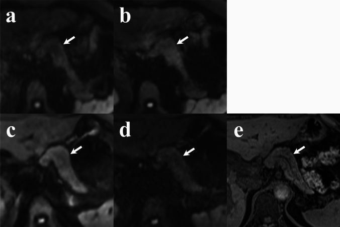
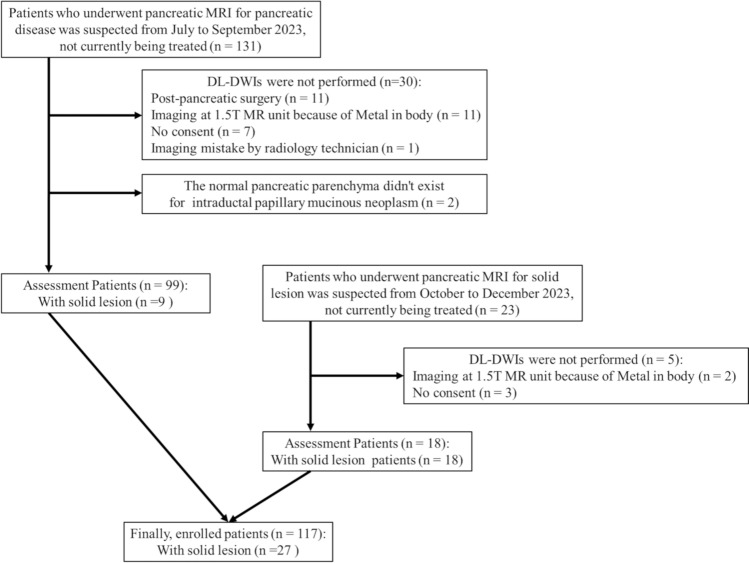
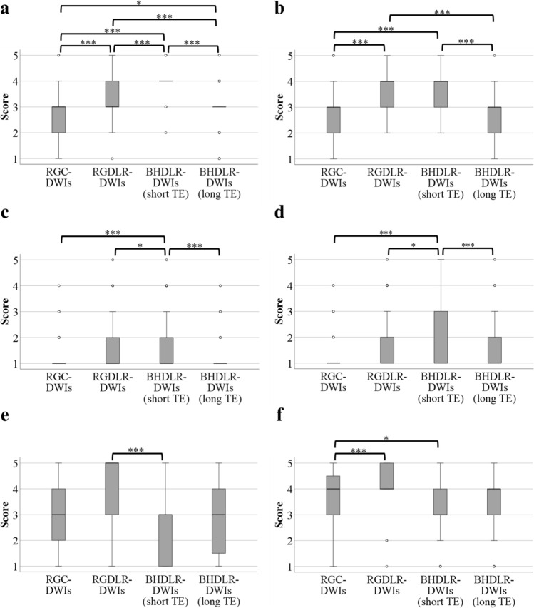
Materials and methods: In total, 117 patients (mean age of 68.0 ± 12.9 years) suspected of pancreatic diseases underwent magnetic resonance imaging (MRI) between July and December 2023. MRI sequences included respiratory-gated conventional diffusion-weighted images (RGC-DWIs), respiratory-gated diffusion-weighted images with deep learning-based reconstruction (DLR) (RGDLR-DWIs), and breath-hold diffusion-weighted images with DLR (BHDLR-DWIs) (short TE and long TE equal to other DWIs) at a 3 T MR system. Among these patients, 27 had solid lesions. Two radiologists qualitatively assessed pancreatic shape, main pancreatic duct (MPD) visualization, and solid lesion conspicuity using a 5-point scale. Quantitative analysis included apparent diffusion coefficient (ADC) values for pancreatic parenchyma and solid lesions, signal-to-noise ratio (SNR), pancreas-to-muscle signal-intensity ratio (PM-SIR) and lesion-to-pancreas signal-intensity ratio (LP-SIR). Differences among DWI sequences were analyzed using Friedman's and Bonferroni's tests.
Results: Qualitatively, BHDLR-DWIs (short TE) had the highest scores for pancreatic shape and MPD but lowest for solid lesions visibility, whereas RGDLR-DWIs had the highest score for solid lesions. Quantitatively, BHDLR-DWIs (short TE) had the lowest ADC values for pancreatic parenchyma and solid lesions, with the highest PM-SIR. There was no significant difference between BHDLR-DWIs (short TE) and RGDLR-DWIs for solid lesion ADC values. RGC-DWIs had the highest SNR, though differences from RGDLR-DWIs and BHDLR-DWIs (short TE) were not significant. Although LP-SIR in RGDLR-DWIs were the lowest, the difference was not significant.
Conclusion: BHDLR-DWIs (short TE) provided the best pancreatic morphology image quality, whereas RGDLR-DWIs were superior for solid lesion detection.
期刊介绍:
Japanese Journal of Radiology is a peer-reviewed journal, officially published by the Japan Radiological Society. The main purpose of the journal is to provide a forum for the publication of papers documenting recent advances and new developments in the field of radiology in medicine and biology. The scope of Japanese Journal of Radiology encompasses but is not restricted to diagnostic radiology, interventional radiology, radiation oncology, nuclear medicine, radiation physics, and radiation biology. Additionally, the journal covers technical and industrial innovations. The journal welcomes original articles, technical notes, review articles, pictorial essays and letters to the editor. The journal also provides announcements from the boards and the committees of the society. Membership in the Japan Radiological Society is not a prerequisite for submission. Contributions are welcomed from all parts of the world.

 求助内容:
求助内容: 应助结果提醒方式:


