Christian Etzold, Orestis Lyros, Matthias Mehdorn, Robert Nowotny, Stefan Niebisch, Boris Jansen-Winkeln, Katrin Schierle, Ines Gockel, René Thieme
{"title":"来自腹膜转移的患者特异性3d组织切片-个体易感性分析的体外模型。","authors":"Christian Etzold, Orestis Lyros, Matthias Mehdorn, Robert Nowotny, Stefan Niebisch, Boris Jansen-Winkeln, Katrin Schierle, Ines Gockel, René Thieme","doi":"10.1515/pp-2024-0012","DOIUrl":null,"url":null,"abstract":"<p><strong>Objectives: </strong>The prognosis of patients with peritoneal metastases (PM) is poor, and these patients have a brief overall survival. Most patients with advanced PM receive palliative therapy to maintain their quality of life. In our current study, we investigated whether patient-specific 3D-tissue slices from patients with PM subjected to pressurized intraperitoneal aerosol chemotherapy could be cultured <i>in vitro</i>.</p><p><strong>Methods: </strong>Biopsies from gastric cancer patients with PM were characterized for cytokeratin-positive tumor cells and the proliferation marker Ki-67. Biopsies from seven patients were cut to 350 µM thick slices in a standardized manner, cultured with 10 µM 5-fluorouracil, doxorubicin, cisplatin, oxaliplatin, or irinotecan for 96 h, and then examined histopathologically and via immunohistochemistry for persistent cytokeratin and Ki-67 expression.</p><p><strong>Results: </strong><i>In vitro</i> cultured slices revealed a similar morphology to un-cultured specimens, and Ki-67-positive tumor cell areas were present after 96 h. The total amount of tumor cells per slice was determined by pan-cytokeratin staining. In the doxorubicin-treated slices, the cytokeratin-positive tumor cell fraction and proliferative (Ki-67pos) cells were decreased. Patient-specific 3D-tissue-slice cultures from peritoneal biopsies were cultured <i>in vitro</i> for up to 4 days.</p><p><strong>Conclusions: </strong>Potentially, these cultures are a reliable model to evaluate the chemosensitivity of patients with PM. Further investigation is needed to match the chemosensitivity with the clinical course of these patients.</p>","PeriodicalId":20231,"journal":{"name":"Pleura and Peritoneum","volume":"10 1","pages":"1-9"},"PeriodicalIF":2.4000,"publicationDate":"2025-02-26","publicationTypes":"Journal Article","fieldsOfStudy":null,"isOpenAccess":false,"openAccessPdf":"https://www.ncbi.nlm.nih.gov/pmc/articles/PMC12016018/pdf/","citationCount":"0","resultStr":"{\"title\":\"Patient-specific 3D-tissue slices from peritoneal metastases - An <i>in vitro</i> model for individual susceptibility analysis.\",\"authors\":\"Christian Etzold, Orestis Lyros, Matthias Mehdorn, Robert Nowotny, Stefan Niebisch, Boris Jansen-Winkeln, Katrin Schierle, Ines Gockel, René Thieme\",\"doi\":\"10.1515/pp-2024-0012\",\"DOIUrl\":null,\"url\":null,\"abstract\":\"<p><strong>Objectives: </strong>The prognosis of patients with peritoneal metastases (PM) is poor, and these patients have a brief overall survival. Most patients with advanced PM receive palliative therapy to maintain their quality of life. In our current study, we investigated whether patient-specific 3D-tissue slices from patients with PM subjected to pressurized intraperitoneal aerosol chemotherapy could be cultured <i>in vitro</i>.</p><p><strong>Methods: </strong>Biopsies from gastric cancer patients with PM were characterized for cytokeratin-positive tumor cells and the proliferation marker Ki-67. Biopsies from seven patients were cut to 350 µM thick slices in a standardized manner, cultured with 10 µM 5-fluorouracil, doxorubicin, cisplatin, oxaliplatin, or irinotecan for 96 h, and then examined histopathologically and via immunohistochemistry for persistent cytokeratin and Ki-67 expression.</p><p><strong>Results: </strong><i>In vitro</i> cultured slices revealed a similar morphology to un-cultured specimens, and Ki-67-positive tumor cell areas were present after 96 h. The total amount of tumor cells per slice was determined by pan-cytokeratin staining. In the doxorubicin-treated slices, the cytokeratin-positive tumor cell fraction and proliferative (Ki-67pos) cells were decreased. Patient-specific 3D-tissue-slice cultures from peritoneal biopsies were cultured <i>in vitro</i> for up to 4 days.</p><p><strong>Conclusions: </strong>Potentially, these cultures are a reliable model to evaluate the chemosensitivity of patients with PM. Further investigation is needed to match the chemosensitivity with the clinical course of these patients.</p>\",\"PeriodicalId\":20231,\"journal\":{\"name\":\"Pleura and Peritoneum\",\"volume\":\"10 1\",\"pages\":\"1-9\"},\"PeriodicalIF\":2.4000,\"publicationDate\":\"2025-02-26\",\"publicationTypes\":\"Journal Article\",\"fieldsOfStudy\":null,\"isOpenAccess\":false,\"openAccessPdf\":\"https://www.ncbi.nlm.nih.gov/pmc/articles/PMC12016018/pdf/\",\"citationCount\":\"0\",\"resultStr\":null,\"platform\":\"Semanticscholar\",\"paperid\":null,\"PeriodicalName\":\"Pleura and Peritoneum\",\"FirstCategoryId\":\"1085\",\"ListUrlMain\":\"https://doi.org/10.1515/pp-2024-0012\",\"RegionNum\":0,\"RegionCategory\":null,\"ArticlePicture\":[],\"TitleCN\":null,\"AbstractTextCN\":null,\"PMCID\":null,\"EPubDate\":\"2025/3/1 0:00:00\",\"PubModel\":\"eCollection\",\"JCR\":\"Q4\",\"JCRName\":\"ONCOLOGY\",\"Score\":null,\"Total\":0}","platform":"Semanticscholar","paperid":null,"PeriodicalName":"Pleura and Peritoneum","FirstCategoryId":"1085","ListUrlMain":"https://doi.org/10.1515/pp-2024-0012","RegionNum":0,"RegionCategory":null,"ArticlePicture":[],"TitleCN":null,"AbstractTextCN":null,"PMCID":null,"EPubDate":"2025/3/1 0:00:00","PubModel":"eCollection","JCR":"Q4","JCRName":"ONCOLOGY","Score":null,"Total":0}
Patient-specific 3D-tissue slices from peritoneal metastases - An in vitro model for individual susceptibility analysis.
Objectives: The prognosis of patients with peritoneal metastases (PM) is poor, and these patients have a brief overall survival. Most patients with advanced PM receive palliative therapy to maintain their quality of life. In our current study, we investigated whether patient-specific 3D-tissue slices from patients with PM subjected to pressurized intraperitoneal aerosol chemotherapy could be cultured in vitro.
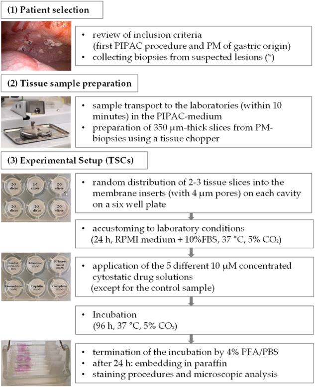
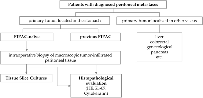
Methods: Biopsies from gastric cancer patients with PM were characterized for cytokeratin-positive tumor cells and the proliferation marker Ki-67. Biopsies from seven patients were cut to 350 µM thick slices in a standardized manner, cultured with 10 µM 5-fluorouracil, doxorubicin, cisplatin, oxaliplatin, or irinotecan for 96 h, and then examined histopathologically and via immunohistochemistry for persistent cytokeratin and Ki-67 expression.
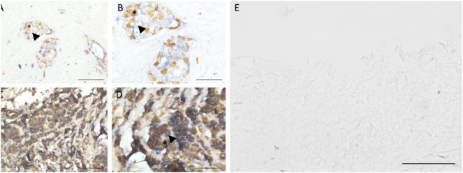
Results: In vitro cultured slices revealed a similar morphology to un-cultured specimens, and Ki-67-positive tumor cell areas were present after 96 h. The total amount of tumor cells per slice was determined by pan-cytokeratin staining. In the doxorubicin-treated slices, the cytokeratin-positive tumor cell fraction and proliferative (Ki-67pos) cells were decreased. Patient-specific 3D-tissue-slice cultures from peritoneal biopsies were cultured in vitro for up to 4 days.
Conclusions: Potentially, these cultures are a reliable model to evaluate the chemosensitivity of patients with PM. Further investigation is needed to match the chemosensitivity with the clinical course of these patients.

 求助内容:
求助内容: 应助结果提醒方式:


