Laurentia Schuster, Sonja Sielker, Johannes Kleinheinz, Till Dammaschke
{"title":"光固化盖髓材料对体外人牙髓细胞的影响。","authors":"Laurentia Schuster, Sonja Sielker, Johannes Kleinheinz, Till Dammaschke","doi":"10.1111/iej.14242","DOIUrl":null,"url":null,"abstract":"<div>\n \n \n <section>\n \n <h3> Aim</h3>\n \n <p>Gold standard as material in vital pulp therapy (VPT) is hydraulic calcium silicate cements (HCSC). To circumvent their prolonged setting time, light-cured pulp capping materials containing calcium silicate or calcium hydroxide powder are available. Although their positive biological properties are advertised, the data regarding the biocompatibility of light-cured pulp capping materials (LCPCM) is inconclusive. This in vitro study compared the biocompatibility of five LCPCM containing calcium silicate (TheraCal LC, ReviCal, MTA PulpCap, Pulprotec MTA) or calcium hydroxide (Calcimol LC) with that of the HCSC Biodentine.</p>\n </section>\n \n <section>\n \n <h3> Methods</h3>\n \n <p>Of each material, 226 cylindrical specimens (51.472 mm<sup>3</sup>) were prepared and incubated in a sterile cell culture medium (alpha Modified Eagle Medium) for 24 h to obtain an extract. Human dental pulp cells (hDPC) were added to the specimens and/or extracts. Cell viability and changes in cell morphology were examined (MTT, LDH, live-dead staining, light microscope). Calcium release from the materials (Ca<sup>2+</sup> colorimetric assay) and the mineralisation capacity of the cells (Alizarin Red S Staining, Alkaline Phosphatase Assay) were determined. Statistical analysis was performed by <span>anova</span> and the post-hoc Tukey test (<i>p</i> < 0.05).</p>\n </section>\n \n <section>\n \n <h3> Results</h3>\n \n <p>Compared to Biodentine, hDPC showed significantly lower cell viability when in contact with LCPCM (<i>p</i> < 0.05). Further, an inhibition zone around the test bodies or an altered cell morphology was observed. Biodentine showed almost no negative effects on cell viability or cell morphology. In contact with Biodentine, hDPC mineralise with and without mineralisation induction conditions. Among the LCPCM, mineralisation was only detectable under induction conditions with ReViCal and MTA PulpCap. In addition, Biodentine released significantly more calcium ions than the LCPCM (<i>p</i> < 0.05).</p>\n </section>\n \n <section>\n \n <h3> Conclusion</h3>\n \n <p>In this in vitro study, LCPCM showed cytotoxic effects on hDPC and were hardly able to induce cell mineralisation. Biodentine showed little negative effects on cell viability, induced cell mineralisation and released more calcium than LCPCM. Biodentine is significantly superior to LCPCM in terms of biocompatibility and mineralisation induction capacity.</p>\n </section>\n </div>","PeriodicalId":13724,"journal":{"name":"International endodontic journal","volume":"58 7","pages":"1060-1072"},"PeriodicalIF":7.1000,"publicationDate":"2025-04-25","publicationTypes":"Journal Article","fieldsOfStudy":null,"isOpenAccess":false,"openAccessPdf":"https://onlinelibrary.wiley.com/doi/epdf/10.1111/iej.14242","citationCount":"0","resultStr":"{\"title\":\"Effect of light-cured pulp capping materials on human dental pulp cells in vitro\",\"authors\":\"Laurentia Schuster, Sonja Sielker, Johannes Kleinheinz, Till Dammaschke\",\"doi\":\"10.1111/iej.14242\",\"DOIUrl\":null,\"url\":null,\"abstract\":\"<div>\\n \\n \\n <section>\\n \\n <h3> Aim</h3>\\n \\n <p>Gold standard as material in vital pulp therapy (VPT) is hydraulic calcium silicate cements (HCSC). To circumvent their prolonged setting time, light-cured pulp capping materials containing calcium silicate or calcium hydroxide powder are available. Although their positive biological properties are advertised, the data regarding the biocompatibility of light-cured pulp capping materials (LCPCM) is inconclusive. This in vitro study compared the biocompatibility of five LCPCM containing calcium silicate (TheraCal LC, ReviCal, MTA PulpCap, Pulprotec MTA) or calcium hydroxide (Calcimol LC) with that of the HCSC Biodentine.</p>\\n </section>\\n \\n <section>\\n \\n <h3> Methods</h3>\\n \\n <p>Of each material, 226 cylindrical specimens (51.472 mm<sup>3</sup>) were prepared and incubated in a sterile cell culture medium (alpha Modified Eagle Medium) for 24 h to obtain an extract. Human dental pulp cells (hDPC) were added to the specimens and/or extracts. Cell viability and changes in cell morphology were examined (MTT, LDH, live-dead staining, light microscope). Calcium release from the materials (Ca<sup>2+</sup> colorimetric assay) and the mineralisation capacity of the cells (Alizarin Red S Staining, Alkaline Phosphatase Assay) were determined. Statistical analysis was performed by <span>anova</span> and the post-hoc Tukey test (<i>p</i> < 0.05).</p>\\n </section>\\n \\n <section>\\n \\n <h3> Results</h3>\\n \\n <p>Compared to Biodentine, hDPC showed significantly lower cell viability when in contact with LCPCM (<i>p</i> < 0.05). Further, an inhibition zone around the test bodies or an altered cell morphology was observed. Biodentine showed almost no negative effects on cell viability or cell morphology. In contact with Biodentine, hDPC mineralise with and without mineralisation induction conditions. Among the LCPCM, mineralisation was only detectable under induction conditions with ReViCal and MTA PulpCap. In addition, Biodentine released significantly more calcium ions than the LCPCM (<i>p</i> < 0.05).</p>\\n </section>\\n \\n <section>\\n \\n <h3> Conclusion</h3>\\n \\n <p>In this in vitro study, LCPCM showed cytotoxic effects on hDPC and were hardly able to induce cell mineralisation. Biodentine showed little negative effects on cell viability, induced cell mineralisation and released more calcium than LCPCM. Biodentine is significantly superior to LCPCM in terms of biocompatibility and mineralisation induction capacity.</p>\\n </section>\\n </div>\",\"PeriodicalId\":13724,\"journal\":{\"name\":\"International endodontic journal\",\"volume\":\"58 7\",\"pages\":\"1060-1072\"},\"PeriodicalIF\":7.1000,\"publicationDate\":\"2025-04-25\",\"publicationTypes\":\"Journal Article\",\"fieldsOfStudy\":null,\"isOpenAccess\":false,\"openAccessPdf\":\"https://onlinelibrary.wiley.com/doi/epdf/10.1111/iej.14242\",\"citationCount\":\"0\",\"resultStr\":null,\"platform\":\"Semanticscholar\",\"paperid\":null,\"PeriodicalName\":\"International endodontic journal\",\"FirstCategoryId\":\"3\",\"ListUrlMain\":\"https://onlinelibrary.wiley.com/doi/10.1111/iej.14242\",\"RegionNum\":1,\"RegionCategory\":\"医学\",\"ArticlePicture\":[],\"TitleCN\":null,\"AbstractTextCN\":null,\"PMCID\":null,\"EPubDate\":\"\",\"PubModel\":\"\",\"JCR\":\"Q1\",\"JCRName\":\"DENTISTRY, ORAL SURGERY & MEDICINE\",\"Score\":null,\"Total\":0}","platform":"Semanticscholar","paperid":null,"PeriodicalName":"International endodontic journal","FirstCategoryId":"3","ListUrlMain":"https://onlinelibrary.wiley.com/doi/10.1111/iej.14242","RegionNum":1,"RegionCategory":"医学","ArticlePicture":[],"TitleCN":null,"AbstractTextCN":null,"PMCID":null,"EPubDate":"","PubModel":"","JCR":"Q1","JCRName":"DENTISTRY, ORAL SURGERY & MEDICINE","Score":null,"Total":0}
Effect of light-cured pulp capping materials on human dental pulp cells in vitro
Aim
Gold standard as material in vital pulp therapy (VPT) is hydraulic calcium silicate cements (HCSC). To circumvent their prolonged setting time, light-cured pulp capping materials containing calcium silicate or calcium hydroxide powder are available. Although their positive biological properties are advertised, the data regarding the biocompatibility of light-cured pulp capping materials (LCPCM) is inconclusive. This in vitro study compared the biocompatibility of five LCPCM containing calcium silicate (TheraCal LC, ReviCal, MTA PulpCap, Pulprotec MTA) or calcium hydroxide (Calcimol LC) with that of the HCSC Biodentine.
Methods
Of each material, 226 cylindrical specimens (51.472 mm3) were prepared and incubated in a sterile cell culture medium (alpha Modified Eagle Medium) for 24 h to obtain an extract. Human dental pulp cells (hDPC) were added to the specimens and/or extracts. Cell viability and changes in cell morphology were examined (MTT, LDH, live-dead staining, light microscope). Calcium release from the materials (Ca2+ colorimetric assay) and the mineralisation capacity of the cells (Alizarin Red S Staining, Alkaline Phosphatase Assay) were determined. Statistical analysis was performed by anova and the post-hoc Tukey test (p < 0.05).
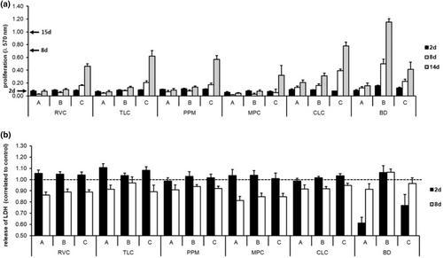
Results
Compared to Biodentine, hDPC showed significantly lower cell viability when in contact with LCPCM (p < 0.05). Further, an inhibition zone around the test bodies or an altered cell morphology was observed. Biodentine showed almost no negative effects on cell viability or cell morphology. In contact with Biodentine, hDPC mineralise with and without mineralisation induction conditions. Among the LCPCM, mineralisation was only detectable under induction conditions with ReViCal and MTA PulpCap. In addition, Biodentine released significantly more calcium ions than the LCPCM (p < 0.05).
Conclusion
In this in vitro study, LCPCM showed cytotoxic effects on hDPC and were hardly able to induce cell mineralisation. Biodentine showed little negative effects on cell viability, induced cell mineralisation and released more calcium than LCPCM. Biodentine is significantly superior to LCPCM in terms of biocompatibility and mineralisation induction capacity.
期刊介绍:
The International Endodontic Journal is published monthly and strives to publish original articles of the highest quality to disseminate scientific and clinical knowledge; all manuscripts are subjected to peer review. Original scientific articles are published in the areas of biomedical science, applied materials science, bioengineering, epidemiology and social science relevant to endodontic disease and its management, and to the restoration of root-treated teeth. In addition, review articles, reports of clinical cases, book reviews, summaries and abstracts of scientific meetings and news items are accepted.
The International Endodontic Journal is essential reading for general dental practitioners, specialist endodontists, research, scientists and dental teachers.

 求助内容:
求助内容: 应助结果提醒方式:


