Soomin Ahn, Yiyu Hong, Sujin Park, Yunjoo Cho, Inwoo Hwang, Ji Min Na, Hyuk Lee, Byung-Hoon Min, Jun Haeng Lee, Jae J Kim, Kyoung-Mee Kim
{"title":"基于深度学习的胃内镜下粘膜剥离标本病理诊断的发展与应用。","authors":"Soomin Ahn, Yiyu Hong, Sujin Park, Yunjoo Cho, Inwoo Hwang, Ji Min Na, Hyuk Lee, Byung-Hoon Min, Jun Haeng Lee, Jae J Kim, Kyoung-Mee Kim","doi":"10.1007/s10120-025-01612-y","DOIUrl":null,"url":null,"abstract":"<p><strong>Background: </strong>Accurate diagnosis of ESD specimens is crucial for managing early gastric cancer. Identifying tumor areas in serially sectioned ESD specimens requires experience and is time-consuming. This study aimed to develop and evaluate a deep learning model for diagnosing ESD specimens.</p><p><strong>Methods: </strong>Whole-slide images of 366 ESD specimens of adenocarcinoma were analyzed, with 2257 annotated regions of interest (tumor and muscularis mucosa) and 83,839 patch images. The development set was divided into training and internal validation sets. Tissue segmentation performance was evaluated using the internal validation set. A detection algorithm for tumor and submucosal invasion at the whole-slide image level was developed, and its performance was evaluated using a test set.</p><p><strong>Results: </strong>The model achieved Dice coefficients of 0.85 and 0.79 for segmentation of tumor and muscularis mucosa, respectively. In the test set, the diagnostic performance of tumor detection, measured by the AUROC, was 0.995, with a specificity of 1.000 and a sensitivity of 0.947. For detecting submucosal invasion, the model achieved an AUROC of 0.981, with a specificity of 0.956 and a sensitivity of 0.907. Pathologists' performance in diagnosing ESD specimens was evaluated with and without assistance from the deep learning model, and the model significantly reduced the mean diagnosis time (747 s without assistance vs. 478 s with assistance, P < 0.001).</p><p><strong>Conclusion: </strong>The deep learning model demonstrated satisfactory performance in tissue segmentation and high accuracy in detecting tumors and submucosal invasion. This model can potentially serve as a screening tool in the histopathological diagnosis of ESD specimens.</p>","PeriodicalId":12684,"journal":{"name":"Gastric Cancer","volume":" ","pages":"609-619"},"PeriodicalIF":5.1000,"publicationDate":"2025-07-01","publicationTypes":"Journal Article","fieldsOfStudy":null,"isOpenAccess":false,"openAccessPdf":"https://www.ncbi.nlm.nih.gov/pmc/articles/PMC12174210/pdf/","citationCount":"0","resultStr":"{\"title\":\"Development and application of deep learning-based diagnostics for pathologic diagnosis of gastric endoscopic submucosal dissection specimens.\",\"authors\":\"Soomin Ahn, Yiyu Hong, Sujin Park, Yunjoo Cho, Inwoo Hwang, Ji Min Na, Hyuk Lee, Byung-Hoon Min, Jun Haeng Lee, Jae J Kim, Kyoung-Mee Kim\",\"doi\":\"10.1007/s10120-025-01612-y\",\"DOIUrl\":null,\"url\":null,\"abstract\":\"<p><strong>Background: </strong>Accurate diagnosis of ESD specimens is crucial for managing early gastric cancer. Identifying tumor areas in serially sectioned ESD specimens requires experience and is time-consuming. This study aimed to develop and evaluate a deep learning model for diagnosing ESD specimens.</p><p><strong>Methods: </strong>Whole-slide images of 366 ESD specimens of adenocarcinoma were analyzed, with 2257 annotated regions of interest (tumor and muscularis mucosa) and 83,839 patch images. The development set was divided into training and internal validation sets. Tissue segmentation performance was evaluated using the internal validation set. A detection algorithm for tumor and submucosal invasion at the whole-slide image level was developed, and its performance was evaluated using a test set.</p><p><strong>Results: </strong>The model achieved Dice coefficients of 0.85 and 0.79 for segmentation of tumor and muscularis mucosa, respectively. In the test set, the diagnostic performance of tumor detection, measured by the AUROC, was 0.995, with a specificity of 1.000 and a sensitivity of 0.947. For detecting submucosal invasion, the model achieved an AUROC of 0.981, with a specificity of 0.956 and a sensitivity of 0.907. Pathologists' performance in diagnosing ESD specimens was evaluated with and without assistance from the deep learning model, and the model significantly reduced the mean diagnosis time (747 s without assistance vs. 478 s with assistance, P < 0.001).</p><p><strong>Conclusion: </strong>The deep learning model demonstrated satisfactory performance in tissue segmentation and high accuracy in detecting tumors and submucosal invasion. This model can potentially serve as a screening tool in the histopathological diagnosis of ESD specimens.</p>\",\"PeriodicalId\":12684,\"journal\":{\"name\":\"Gastric Cancer\",\"volume\":\" \",\"pages\":\"609-619\"},\"PeriodicalIF\":5.1000,\"publicationDate\":\"2025-07-01\",\"publicationTypes\":\"Journal Article\",\"fieldsOfStudy\":null,\"isOpenAccess\":false,\"openAccessPdf\":\"https://www.ncbi.nlm.nih.gov/pmc/articles/PMC12174210/pdf/\",\"citationCount\":\"0\",\"resultStr\":null,\"platform\":\"Semanticscholar\",\"paperid\":null,\"PeriodicalName\":\"Gastric Cancer\",\"FirstCategoryId\":\"3\",\"ListUrlMain\":\"https://doi.org/10.1007/s10120-025-01612-y\",\"RegionNum\":1,\"RegionCategory\":\"医学\",\"ArticlePicture\":[],\"TitleCN\":null,\"AbstractTextCN\":null,\"PMCID\":null,\"EPubDate\":\"2025/4/15 0:00:00\",\"PubModel\":\"Epub\",\"JCR\":\"Q1\",\"JCRName\":\"GASTROENTEROLOGY & HEPATOLOGY\",\"Score\":null,\"Total\":0}","platform":"Semanticscholar","paperid":null,"PeriodicalName":"Gastric Cancer","FirstCategoryId":"3","ListUrlMain":"https://doi.org/10.1007/s10120-025-01612-y","RegionNum":1,"RegionCategory":"医学","ArticlePicture":[],"TitleCN":null,"AbstractTextCN":null,"PMCID":null,"EPubDate":"2025/4/15 0:00:00","PubModel":"Epub","JCR":"Q1","JCRName":"GASTROENTEROLOGY & HEPATOLOGY","Score":null,"Total":0}
Development and application of deep learning-based diagnostics for pathologic diagnosis of gastric endoscopic submucosal dissection specimens.
Background: Accurate diagnosis of ESD specimens is crucial for managing early gastric cancer. Identifying tumor areas in serially sectioned ESD specimens requires experience and is time-consuming. This study aimed to develop and evaluate a deep learning model for diagnosing ESD specimens.
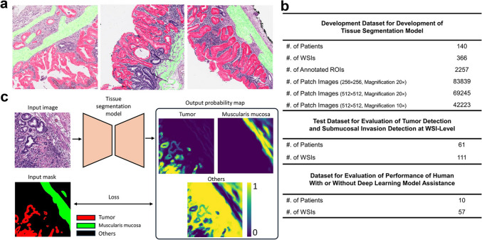
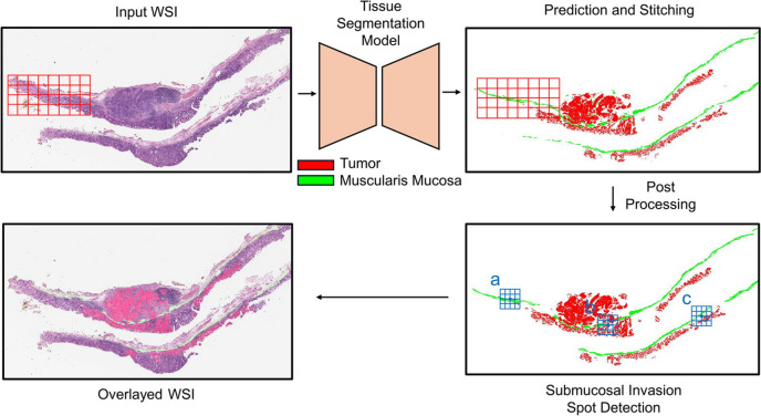
Methods: Whole-slide images of 366 ESD specimens of adenocarcinoma were analyzed, with 2257 annotated regions of interest (tumor and muscularis mucosa) and 83,839 patch images. The development set was divided into training and internal validation sets. Tissue segmentation performance was evaluated using the internal validation set. A detection algorithm for tumor and submucosal invasion at the whole-slide image level was developed, and its performance was evaluated using a test set.
Results: The model achieved Dice coefficients of 0.85 and 0.79 for segmentation of tumor and muscularis mucosa, respectively. In the test set, the diagnostic performance of tumor detection, measured by the AUROC, was 0.995, with a specificity of 1.000 and a sensitivity of 0.947. For detecting submucosal invasion, the model achieved an AUROC of 0.981, with a specificity of 0.956 and a sensitivity of 0.907. Pathologists' performance in diagnosing ESD specimens was evaluated with and without assistance from the deep learning model, and the model significantly reduced the mean diagnosis time (747 s without assistance vs. 478 s with assistance, P < 0.001).
Conclusion: The deep learning model demonstrated satisfactory performance in tissue segmentation and high accuracy in detecting tumors and submucosal invasion. This model can potentially serve as a screening tool in the histopathological diagnosis of ESD specimens.
期刊介绍:
Gastric Cancer is an esteemed global forum that focuses on various aspects of gastric cancer research, treatment, and biology worldwide.
The journal promotes a diverse range of content, including original articles, case reports, short communications, and technical notes. It also welcomes Letters to the Editor discussing published articles or sharing viewpoints on gastric cancer topics.
Review articles are predominantly sought after by the Editor, ensuring comprehensive coverage of the field.
With a dedicated and knowledgeable editorial team, the journal is committed to providing exceptional support and ensuring high levels of author satisfaction. In fact, over 90% of published authors have expressed their intent to publish again in our esteemed journal.
 求助内容:
求助内容: 应助结果提醒方式:


