María Belén Zanchetta, Fernando Jerkovich, Florencia Scioscia, Yamile Mocarbel, Analía Pignatta, Natalia Elías, Juan Manuel Roganovich, Carlos Vigovich, María Celeste Balonga, Ana Carolina Cohen, Giselle Mumbach, José Luis Mansur, Carolina Fux Otta, Walter Guillermo Douthat, Pilar Tartaglia, Griselda Cecchi, María Bastianello, Luisa Plantalech, Erich Fradinger, José Rubén Zanchetta
{"title":"肿瘤性骨软化患者的骨是如何恢复的?一项国家队列研究的长期随访。","authors":"María Belén Zanchetta, Fernando Jerkovich, Florencia Scioscia, Yamile Mocarbel, Analía Pignatta, Natalia Elías, Juan Manuel Roganovich, Carlos Vigovich, María Celeste Balonga, Ana Carolina Cohen, Giselle Mumbach, José Luis Mansur, Carolina Fux Otta, Walter Guillermo Douthat, Pilar Tartaglia, Griselda Cecchi, María Bastianello, Luisa Plantalech, Erich Fradinger, José Rubén Zanchetta","doi":"10.1093/jbmrpl/ziaf041","DOIUrl":null,"url":null,"abstract":"<p><p>Tumor-induced osteomalacia (TIO) is a rare disorder characterized by impaired bone mineralization due to phosphate wasting. Long-term changes in BMD and microarchitecture after surgical cure or medical therapy in TIO are not well understood. This study describes changes in BMD, microarchitecture, and bone strength in patients with TIO following surgical cure or medical therapy. A prospective cohort study included adults diagnosed with TIO from May 2018 to 2024, categorized into those with surgical cure and those on medical therapy. Follow-up assessments were classified as early (median 8 mo), intermediate (median 17 mo), and long-term (median 26 mo). Fifteen patients were included: seven achieved surgical cure, and eight remained on medical therapy. Lumbar spine BMD increased by +19% at early, +27% at intermediate, and +15% at long-term follow-up. Total hip BMD increased by +31%, +36%, and +31% at early, intermediate, and long-term assessments, respectively. All patients achieved a normal lumbar spine BMD, while 91% attained a normal total hip BMD. At the distal tibia, substantial increases in bone microarchitecture parameters-cortical area (Ct.Ar), cortical volumetric density (Ct.vBMD), and cortical thickness (Ct.Th)-were observed. Notably, Ct.Th improved to levels comparable to healthy controls. Bone strength improved by 13% but was not statistically significant, probably due to the small sample size. At the distal radius, most parameters remained stable. Patients with surgical cure showed more rapid and substantial improvements in BMD and cortical microarchitecture than non-cured patients, but these differences did not reach statistical significance. Overall, bone recovery in TIO is gradual, with gains in spine and hip BMD and significant improvements in tibial cortical parameters. However, some aspects of bone microarchitecture remained below control levels, underscoring the need for ongoing monitoring and individualized management strategies.</p>","PeriodicalId":14611,"journal":{"name":"JBMR Plus","volume":"9 5","pages":"ziaf041"},"PeriodicalIF":2.4000,"publicationDate":"2025-04-14","publicationTypes":"Journal Article","fieldsOfStudy":null,"isOpenAccess":false,"openAccessPdf":"https://www.ncbi.nlm.nih.gov/pmc/articles/PMC12010155/pdf/","citationCount":"0","resultStr":"{\"title\":\"How does bone recover in patients with tumor-induced osteomalacia? Long-term follow-up in a national cohort study.\",\"authors\":\"María Belén Zanchetta, Fernando Jerkovich, Florencia Scioscia, Yamile Mocarbel, Analía Pignatta, Natalia Elías, Juan Manuel Roganovich, Carlos Vigovich, María Celeste Balonga, Ana Carolina Cohen, Giselle Mumbach, José Luis Mansur, Carolina Fux Otta, Walter Guillermo Douthat, Pilar Tartaglia, Griselda Cecchi, María Bastianello, Luisa Plantalech, Erich Fradinger, José Rubén Zanchetta\",\"doi\":\"10.1093/jbmrpl/ziaf041\",\"DOIUrl\":null,\"url\":null,\"abstract\":\"<p><p>Tumor-induced osteomalacia (TIO) is a rare disorder characterized by impaired bone mineralization due to phosphate wasting. Long-term changes in BMD and microarchitecture after surgical cure or medical therapy in TIO are not well understood. This study describes changes in BMD, microarchitecture, and bone strength in patients with TIO following surgical cure or medical therapy. A prospective cohort study included adults diagnosed with TIO from May 2018 to 2024, categorized into those with surgical cure and those on medical therapy. Follow-up assessments were classified as early (median 8 mo), intermediate (median 17 mo), and long-term (median 26 mo). Fifteen patients were included: seven achieved surgical cure, and eight remained on medical therapy. Lumbar spine BMD increased by +19% at early, +27% at intermediate, and +15% at long-term follow-up. Total hip BMD increased by +31%, +36%, and +31% at early, intermediate, and long-term assessments, respectively. All patients achieved a normal lumbar spine BMD, while 91% attained a normal total hip BMD. At the distal tibia, substantial increases in bone microarchitecture parameters-cortical area (Ct.Ar), cortical volumetric density (Ct.vBMD), and cortical thickness (Ct.Th)-were observed. Notably, Ct.Th improved to levels comparable to healthy controls. Bone strength improved by 13% but was not statistically significant, probably due to the small sample size. At the distal radius, most parameters remained stable. Patients with surgical cure showed more rapid and substantial improvements in BMD and cortical microarchitecture than non-cured patients, but these differences did not reach statistical significance. Overall, bone recovery in TIO is gradual, with gains in spine and hip BMD and significant improvements in tibial cortical parameters. However, some aspects of bone microarchitecture remained below control levels, underscoring the need for ongoing monitoring and individualized management strategies.</p>\",\"PeriodicalId\":14611,\"journal\":{\"name\":\"JBMR Plus\",\"volume\":\"9 5\",\"pages\":\"ziaf041\"},\"PeriodicalIF\":2.4000,\"publicationDate\":\"2025-04-14\",\"publicationTypes\":\"Journal Article\",\"fieldsOfStudy\":null,\"isOpenAccess\":false,\"openAccessPdf\":\"https://www.ncbi.nlm.nih.gov/pmc/articles/PMC12010155/pdf/\",\"citationCount\":\"0\",\"resultStr\":null,\"platform\":\"Semanticscholar\",\"paperid\":null,\"PeriodicalName\":\"JBMR Plus\",\"FirstCategoryId\":\"1085\",\"ListUrlMain\":\"https://doi.org/10.1093/jbmrpl/ziaf041\",\"RegionNum\":0,\"RegionCategory\":null,\"ArticlePicture\":[],\"TitleCN\":null,\"AbstractTextCN\":null,\"PMCID\":null,\"EPubDate\":\"2025/5/1 0:00:00\",\"PubModel\":\"eCollection\",\"JCR\":\"Q2\",\"JCRName\":\"ENDOCRINOLOGY & METABOLISM\",\"Score\":null,\"Total\":0}","platform":"Semanticscholar","paperid":null,"PeriodicalName":"JBMR Plus","FirstCategoryId":"1085","ListUrlMain":"https://doi.org/10.1093/jbmrpl/ziaf041","RegionNum":0,"RegionCategory":null,"ArticlePicture":[],"TitleCN":null,"AbstractTextCN":null,"PMCID":null,"EPubDate":"2025/5/1 0:00:00","PubModel":"eCollection","JCR":"Q2","JCRName":"ENDOCRINOLOGY & METABOLISM","Score":null,"Total":0}
How does bone recover in patients with tumor-induced osteomalacia? Long-term follow-up in a national cohort study.
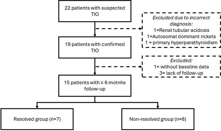
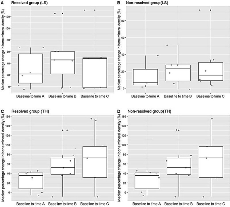
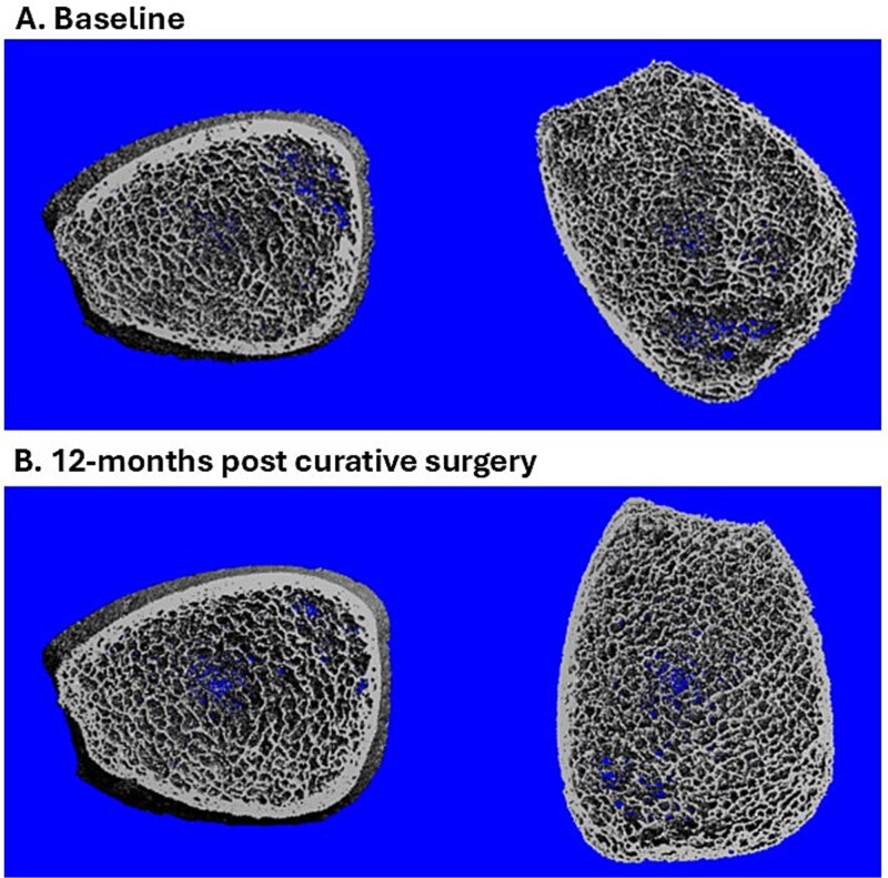
Tumor-induced osteomalacia (TIO) is a rare disorder characterized by impaired bone mineralization due to phosphate wasting. Long-term changes in BMD and microarchitecture after surgical cure or medical therapy in TIO are not well understood. This study describes changes in BMD, microarchitecture, and bone strength in patients with TIO following surgical cure or medical therapy. A prospective cohort study included adults diagnosed with TIO from May 2018 to 2024, categorized into those with surgical cure and those on medical therapy. Follow-up assessments were classified as early (median 8 mo), intermediate (median 17 mo), and long-term (median 26 mo). Fifteen patients were included: seven achieved surgical cure, and eight remained on medical therapy. Lumbar spine BMD increased by +19% at early, +27% at intermediate, and +15% at long-term follow-up. Total hip BMD increased by +31%, +36%, and +31% at early, intermediate, and long-term assessments, respectively. All patients achieved a normal lumbar spine BMD, while 91% attained a normal total hip BMD. At the distal tibia, substantial increases in bone microarchitecture parameters-cortical area (Ct.Ar), cortical volumetric density (Ct.vBMD), and cortical thickness (Ct.Th)-were observed. Notably, Ct.Th improved to levels comparable to healthy controls. Bone strength improved by 13% but was not statistically significant, probably due to the small sample size. At the distal radius, most parameters remained stable. Patients with surgical cure showed more rapid and substantial improvements in BMD and cortical microarchitecture than non-cured patients, but these differences did not reach statistical significance. Overall, bone recovery in TIO is gradual, with gains in spine and hip BMD and significant improvements in tibial cortical parameters. However, some aspects of bone microarchitecture remained below control levels, underscoring the need for ongoing monitoring and individualized management strategies.

 求助内容:
求助内容: 应助结果提醒方式:


