Florence Birru, Anne Hicks, Jake Mandziuk, Ioana Bratu
{"title":"是否有肺气肿?婴儿肺囊性病变病例报告。","authors":"Florence Birru, Anne Hicks, Jake Mandziuk, Ioana Bratu","doi":"10.1177/10935266251338562","DOIUrl":null,"url":null,"abstract":"<p><p>Pulmonary cystic lesions in infants are uncommon and can present diagnostic challenges due to overlapping radiologic features with other cystic lung conditions. We present 2 cases of left lung cystic lesions in infants. Initial high-resolution computed tomography (HRCT) suggested differential diagnoses, including pneumatocele, type I congenital pulmonary airway malformation (CPAM), or a solitary cystic lymphangioma. Definitive diagnosis was achieved through histopathological examination after left lower lobectomy and resection of the lesion. These cases highlight the challenges in accurately diagnosing pulmonary cystic lesions, given the limitations of imaging alone.</p>","PeriodicalId":54634,"journal":{"name":"Pediatric and Developmental Pathology","volume":" ","pages":"400-405"},"PeriodicalIF":1.3000,"publicationDate":"2025-09-01","publicationTypes":"Journal Article","fieldsOfStudy":null,"isOpenAccess":false,"openAccessPdf":"https://www.ncbi.nlm.nih.gov/pmc/articles/PMC12426324/pdf/","citationCount":"0","resultStr":"{\"title\":\"Pneumatoceles or Not? Case Reports on Pulmonary Cystic Lesions in Infants.\",\"authors\":\"Florence Birru, Anne Hicks, Jake Mandziuk, Ioana Bratu\",\"doi\":\"10.1177/10935266251338562\",\"DOIUrl\":null,\"url\":null,\"abstract\":\"<p><p>Pulmonary cystic lesions in infants are uncommon and can present diagnostic challenges due to overlapping radiologic features with other cystic lung conditions. We present 2 cases of left lung cystic lesions in infants. Initial high-resolution computed tomography (HRCT) suggested differential diagnoses, including pneumatocele, type I congenital pulmonary airway malformation (CPAM), or a solitary cystic lymphangioma. Definitive diagnosis was achieved through histopathological examination after left lower lobectomy and resection of the lesion. These cases highlight the challenges in accurately diagnosing pulmonary cystic lesions, given the limitations of imaging alone.</p>\",\"PeriodicalId\":54634,\"journal\":{\"name\":\"Pediatric and Developmental Pathology\",\"volume\":\" \",\"pages\":\"400-405\"},\"PeriodicalIF\":1.3000,\"publicationDate\":\"2025-09-01\",\"publicationTypes\":\"Journal Article\",\"fieldsOfStudy\":null,\"isOpenAccess\":false,\"openAccessPdf\":\"https://www.ncbi.nlm.nih.gov/pmc/articles/PMC12426324/pdf/\",\"citationCount\":\"0\",\"resultStr\":null,\"platform\":\"Semanticscholar\",\"paperid\":null,\"PeriodicalName\":\"Pediatric and Developmental Pathology\",\"FirstCategoryId\":\"3\",\"ListUrlMain\":\"https://doi.org/10.1177/10935266251338562\",\"RegionNum\":4,\"RegionCategory\":\"医学\",\"ArticlePicture\":[],\"TitleCN\":null,\"AbstractTextCN\":null,\"PMCID\":null,\"EPubDate\":\"2025/5/12 0:00:00\",\"PubModel\":\"Epub\",\"JCR\":\"Q3\",\"JCRName\":\"PATHOLOGY\",\"Score\":null,\"Total\":0}","platform":"Semanticscholar","paperid":null,"PeriodicalName":"Pediatric and Developmental Pathology","FirstCategoryId":"3","ListUrlMain":"https://doi.org/10.1177/10935266251338562","RegionNum":4,"RegionCategory":"医学","ArticlePicture":[],"TitleCN":null,"AbstractTextCN":null,"PMCID":null,"EPubDate":"2025/5/12 0:00:00","PubModel":"Epub","JCR":"Q3","JCRName":"PATHOLOGY","Score":null,"Total":0}
Pneumatoceles or Not? Case Reports on Pulmonary Cystic Lesions in Infants.
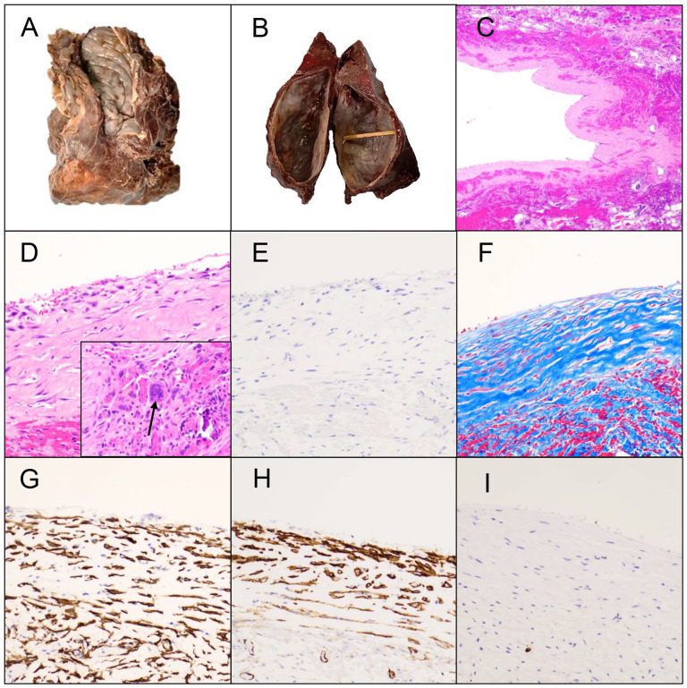
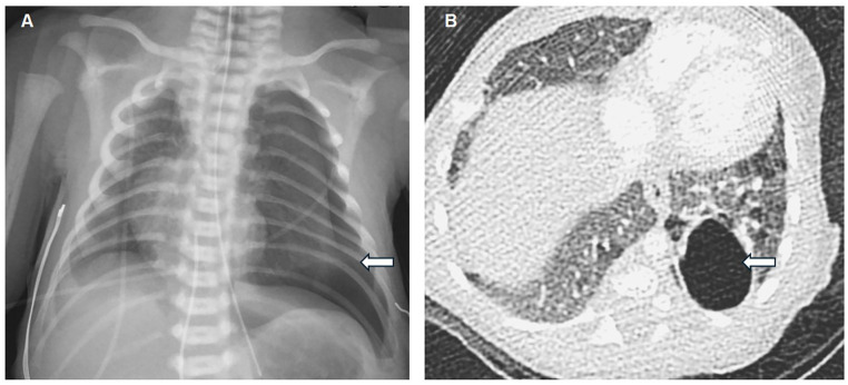
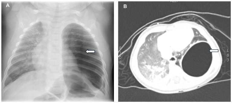
Pulmonary cystic lesions in infants are uncommon and can present diagnostic challenges due to overlapping radiologic features with other cystic lung conditions. We present 2 cases of left lung cystic lesions in infants. Initial high-resolution computed tomography (HRCT) suggested differential diagnoses, including pneumatocele, type I congenital pulmonary airway malformation (CPAM), or a solitary cystic lymphangioma. Definitive diagnosis was achieved through histopathological examination after left lower lobectomy and resection of the lesion. These cases highlight the challenges in accurately diagnosing pulmonary cystic lesions, given the limitations of imaging alone.
期刊介绍:
The Journal covers the spectrum of disorders of early development (including embryology, placentology, and teratology), gestational and perinatal diseases, and all diseases of childhood. Studies may be in any field of experimental, anatomic, or clinical pathology, including molecular pathology. Case reports are published only if they provide new insights into disease mechanisms or new information.
 求助内容:
求助内容: 应助结果提醒方式:


