{"title":"基于多模态磁共振图像诊断脑卒中类型和严重程度的多分类深度学习方法。","authors":"Sahar Felehgari, Payam Sariaslani, Sepideh Shamsizadeh, Saba Felehgari, Anahita Rajabi, Hiwa Mohammadi","doi":"10.4103/jmss.jmss_37_24","DOIUrl":null,"url":null,"abstract":"<p><strong>Background: </strong>Clinical decisions for stroke treatments, such as thrombolytic drugs for ischemic strokes or anticoagulants for hemorrhagic strokes, rely on accurate diagnosis and severity assessment. Our study uses diffusion-weighted magnetic resonance imaging and Convolutional Neural Networks (CNNs) to differentiate healthy and stroke samples, classify stroke types, and predict severity, aiding in decision-making for stroke management.</p><p><strong>Methods: </strong>We evaluated 143 patients: 85 with ischemic stroke and 58 with hemorrhagic stroke. For stroke diagnosis, we compared multimodal (apparent diffusion coefficient and diffusion-weighted imaging [DWI]) and single-modal (using separate images) preprocessing techniques. Our study introduced two models, Added CNN Layer-ResNet-50 (ACL-ResNet-50) and Added CNN Layer-MobileNetV1 (ACL-MobileNetV1), based on transfer learning (MobileNetV1 and ResNet-50), enhancing performance through reinforced layers. We compared our proposed models with a scenario in which only the final layer was replaced in ResNet-50 and MobileNetV1. Furthermore, we predicted National Institutes of Health Stroke Scale (NIHSS) scores in three ranges based on DWI images to gauge stroke severity. Evaluation criteria for the models included accuracy, sensitivity, specificity, and area under the curve (AUC).</p><p><strong>Results: </strong>In stroke classification (normal, ischemic, and hemorrhagic), ACL-MobileNetV1 outperformed other models, achieving 98% accuracy, 99% sensitivity, 98% specificity, and 99% AUC. For assessing ischemic stroke severity using NIHSS ranges, ACL-ResNet-50 showed the optimal performance with an accuracy of 0.92, sensitivity of 0.84, specificity of 0.92, and AUC of 0.95.</p><p><strong>Conclusion: </strong>Our study's proposed method effectively classified stroke type and severity based on multimodal MR images, potentially as a practical decision support tool for stroke treatments.</p>","PeriodicalId":37680,"journal":{"name":"Journal of Medical Signals & Sensors","volume":"15 ","pages":"10"},"PeriodicalIF":1.1000,"publicationDate":"2025-04-19","publicationTypes":"Journal Article","fieldsOfStudy":null,"isOpenAccess":false,"openAccessPdf":"https://www.ncbi.nlm.nih.gov/pmc/articles/PMC12063969/pdf/","citationCount":"0","resultStr":"{\"title\":\"Multi-classification Deep Learning Approach for Diagnosing Stroke Type and Severity Using Multimodal Magnetic Resonance Images.\",\"authors\":\"Sahar Felehgari, Payam Sariaslani, Sepideh Shamsizadeh, Saba Felehgari, Anahita Rajabi, Hiwa Mohammadi\",\"doi\":\"10.4103/jmss.jmss_37_24\",\"DOIUrl\":null,\"url\":null,\"abstract\":\"<p><strong>Background: </strong>Clinical decisions for stroke treatments, such as thrombolytic drugs for ischemic strokes or anticoagulants for hemorrhagic strokes, rely on accurate diagnosis and severity assessment. Our study uses diffusion-weighted magnetic resonance imaging and Convolutional Neural Networks (CNNs) to differentiate healthy and stroke samples, classify stroke types, and predict severity, aiding in decision-making for stroke management.</p><p><strong>Methods: </strong>We evaluated 143 patients: 85 with ischemic stroke and 58 with hemorrhagic stroke. For stroke diagnosis, we compared multimodal (apparent diffusion coefficient and diffusion-weighted imaging [DWI]) and single-modal (using separate images) preprocessing techniques. Our study introduced two models, Added CNN Layer-ResNet-50 (ACL-ResNet-50) and Added CNN Layer-MobileNetV1 (ACL-MobileNetV1), based on transfer learning (MobileNetV1 and ResNet-50), enhancing performance through reinforced layers. We compared our proposed models with a scenario in which only the final layer was replaced in ResNet-50 and MobileNetV1. Furthermore, we predicted National Institutes of Health Stroke Scale (NIHSS) scores in three ranges based on DWI images to gauge stroke severity. Evaluation criteria for the models included accuracy, sensitivity, specificity, and area under the curve (AUC).</p><p><strong>Results: </strong>In stroke classification (normal, ischemic, and hemorrhagic), ACL-MobileNetV1 outperformed other models, achieving 98% accuracy, 99% sensitivity, 98% specificity, and 99% AUC. For assessing ischemic stroke severity using NIHSS ranges, ACL-ResNet-50 showed the optimal performance with an accuracy of 0.92, sensitivity of 0.84, specificity of 0.92, and AUC of 0.95.</p><p><strong>Conclusion: </strong>Our study's proposed method effectively classified stroke type and severity based on multimodal MR images, potentially as a practical decision support tool for stroke treatments.</p>\",\"PeriodicalId\":37680,\"journal\":{\"name\":\"Journal of Medical Signals & Sensors\",\"volume\":\"15 \",\"pages\":\"10\"},\"PeriodicalIF\":1.1000,\"publicationDate\":\"2025-04-19\",\"publicationTypes\":\"Journal Article\",\"fieldsOfStudy\":null,\"isOpenAccess\":false,\"openAccessPdf\":\"https://www.ncbi.nlm.nih.gov/pmc/articles/PMC12063969/pdf/\",\"citationCount\":\"0\",\"resultStr\":null,\"platform\":\"Semanticscholar\",\"paperid\":null,\"PeriodicalName\":\"Journal of Medical Signals & Sensors\",\"FirstCategoryId\":\"1085\",\"ListUrlMain\":\"https://doi.org/10.4103/jmss.jmss_37_24\",\"RegionNum\":0,\"RegionCategory\":null,\"ArticlePicture\":[],\"TitleCN\":null,\"AbstractTextCN\":null,\"PMCID\":null,\"EPubDate\":\"2025/1/1 0:00:00\",\"PubModel\":\"eCollection\",\"JCR\":\"Q4\",\"JCRName\":\"ENGINEERING, BIOMEDICAL\",\"Score\":null,\"Total\":0}","platform":"Semanticscholar","paperid":null,"PeriodicalName":"Journal of Medical Signals & Sensors","FirstCategoryId":"1085","ListUrlMain":"https://doi.org/10.4103/jmss.jmss_37_24","RegionNum":0,"RegionCategory":null,"ArticlePicture":[],"TitleCN":null,"AbstractTextCN":null,"PMCID":null,"EPubDate":"2025/1/1 0:00:00","PubModel":"eCollection","JCR":"Q4","JCRName":"ENGINEERING, BIOMEDICAL","Score":null,"Total":0}
Multi-classification Deep Learning Approach for Diagnosing Stroke Type and Severity Using Multimodal Magnetic Resonance Images.
Background: Clinical decisions for stroke treatments, such as thrombolytic drugs for ischemic strokes or anticoagulants for hemorrhagic strokes, rely on accurate diagnosis and severity assessment. Our study uses diffusion-weighted magnetic resonance imaging and Convolutional Neural Networks (CNNs) to differentiate healthy and stroke samples, classify stroke types, and predict severity, aiding in decision-making for stroke management.
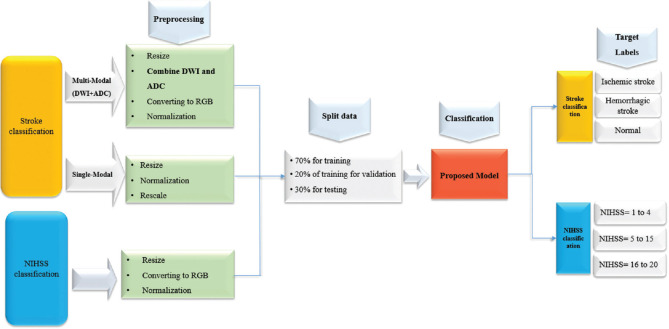
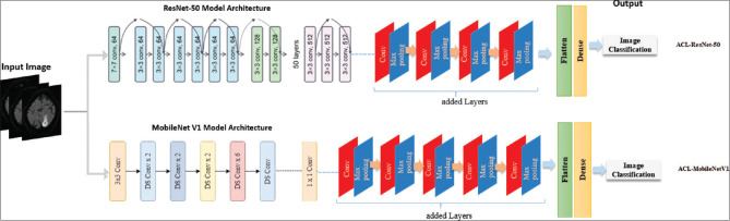
Methods: We evaluated 143 patients: 85 with ischemic stroke and 58 with hemorrhagic stroke. For stroke diagnosis, we compared multimodal (apparent diffusion coefficient and diffusion-weighted imaging [DWI]) and single-modal (using separate images) preprocessing techniques. Our study introduced two models, Added CNN Layer-ResNet-50 (ACL-ResNet-50) and Added CNN Layer-MobileNetV1 (ACL-MobileNetV1), based on transfer learning (MobileNetV1 and ResNet-50), enhancing performance through reinforced layers. We compared our proposed models with a scenario in which only the final layer was replaced in ResNet-50 and MobileNetV1. Furthermore, we predicted National Institutes of Health Stroke Scale (NIHSS) scores in three ranges based on DWI images to gauge stroke severity. Evaluation criteria for the models included accuracy, sensitivity, specificity, and area under the curve (AUC).
Results: In stroke classification (normal, ischemic, and hemorrhagic), ACL-MobileNetV1 outperformed other models, achieving 98% accuracy, 99% sensitivity, 98% specificity, and 99% AUC. For assessing ischemic stroke severity using NIHSS ranges, ACL-ResNet-50 showed the optimal performance with an accuracy of 0.92, sensitivity of 0.84, specificity of 0.92, and AUC of 0.95.
Conclusion: Our study's proposed method effectively classified stroke type and severity based on multimodal MR images, potentially as a practical decision support tool for stroke treatments.
期刊介绍:
JMSS is an interdisciplinary journal that incorporates all aspects of the biomedical engineering including bioelectrics, bioinformatics, medical physics, health technology assessment, etc. Subject areas covered by the journal include: - Bioelectric: Bioinstruments Biosensors Modeling Biomedical signal processing Medical image analysis and processing Medical imaging devices Control of biological systems Neuromuscular systems Cognitive sciences Telemedicine Robotic Medical ultrasonography Bioelectromagnetics Electrophysiology Cell tracking - Bioinformatics and medical informatics: Analysis of biological data Data mining Stochastic modeling Computational genomics Artificial intelligence & fuzzy Applications Medical softwares Bioalgorithms Electronic health - Biophysics and medical physics: Computed tomography Radiation therapy Laser therapy - Education in biomedical engineering - Health technology assessment - Standard in biomedical engineering.

 求助内容:
求助内容: 应助结果提醒方式:


