Anna Pitsillidi, Sergios Ion Karras, Günter Karl Noé
{"title":"左髂外动脉平滑肌肉瘤1例报告及文献回顾。","authors":"Anna Pitsillidi, Sergios Ion Karras, Günter Karl Noé","doi":"10.52054/FVVO.2024.13623","DOIUrl":null,"url":null,"abstract":"<p><p>Leiomyosarcomas (LMS) arise from smooth muscle and represents only 6% of all sarcomas. LMS originating from major blood vessels, called vascular LMS, are detected mostly in the inferior vena cava. Arterial LMS are a rarity. We present a 43-year-old patient with a LMS arising from the left external iliac artery. The patient was referred to us with symptoms of left lower abdominal pain extending to the left limb and underwent a contrast computed tomography which suggested a suspicious mass near the left iliac vessels. She underwent laparoscopic excision of the tumour, whose histological examination revealed an LMS G2 arising from the external iliac artery. Immunohistochemically CD34, p53, Desmin, as well as smooth muscle actin, tested positive.</p>","PeriodicalId":46400,"journal":{"name":"Facts Views and Vision in ObGyn","volume":"17 1","pages":"94-98"},"PeriodicalIF":1.4000,"publicationDate":"2025-03-28","publicationTypes":"Journal Article","fieldsOfStudy":null,"isOpenAccess":false,"openAccessPdf":"https://www.ncbi.nlm.nih.gov/pmc/articles/PMC12042151/pdf/","citationCount":"0","resultStr":"{\"title\":\"Leiomyosarcoma of the left external iliac artery: a case report and narrative review of the literature.\",\"authors\":\"Anna Pitsillidi, Sergios Ion Karras, Günter Karl Noé\",\"doi\":\"10.52054/FVVO.2024.13623\",\"DOIUrl\":null,\"url\":null,\"abstract\":\"<p><p>Leiomyosarcomas (LMS) arise from smooth muscle and represents only 6% of all sarcomas. LMS originating from major blood vessels, called vascular LMS, are detected mostly in the inferior vena cava. Arterial LMS are a rarity. We present a 43-year-old patient with a LMS arising from the left external iliac artery. The patient was referred to us with symptoms of left lower abdominal pain extending to the left limb and underwent a contrast computed tomography which suggested a suspicious mass near the left iliac vessels. She underwent laparoscopic excision of the tumour, whose histological examination revealed an LMS G2 arising from the external iliac artery. Immunohistochemically CD34, p53, Desmin, as well as smooth muscle actin, tested positive.</p>\",\"PeriodicalId\":46400,\"journal\":{\"name\":\"Facts Views and Vision in ObGyn\",\"volume\":\"17 1\",\"pages\":\"94-98\"},\"PeriodicalIF\":1.4000,\"publicationDate\":\"2025-03-28\",\"publicationTypes\":\"Journal Article\",\"fieldsOfStudy\":null,\"isOpenAccess\":false,\"openAccessPdf\":\"https://www.ncbi.nlm.nih.gov/pmc/articles/PMC12042151/pdf/\",\"citationCount\":\"0\",\"resultStr\":null,\"platform\":\"Semanticscholar\",\"paperid\":null,\"PeriodicalName\":\"Facts Views and Vision in ObGyn\",\"FirstCategoryId\":\"1085\",\"ListUrlMain\":\"https://doi.org/10.52054/FVVO.2024.13623\",\"RegionNum\":0,\"RegionCategory\":null,\"ArticlePicture\":[],\"TitleCN\":null,\"AbstractTextCN\":null,\"PMCID\":null,\"EPubDate\":\"\",\"PubModel\":\"\",\"JCR\":\"Q3\",\"JCRName\":\"OBSTETRICS & GYNECOLOGY\",\"Score\":null,\"Total\":0}","platform":"Semanticscholar","paperid":null,"PeriodicalName":"Facts Views and Vision in ObGyn","FirstCategoryId":"1085","ListUrlMain":"https://doi.org/10.52054/FVVO.2024.13623","RegionNum":0,"RegionCategory":null,"ArticlePicture":[],"TitleCN":null,"AbstractTextCN":null,"PMCID":null,"EPubDate":"","PubModel":"","JCR":"Q3","JCRName":"OBSTETRICS & GYNECOLOGY","Score":null,"Total":0}
Leiomyosarcoma of the left external iliac artery: a case report and narrative review of the literature.
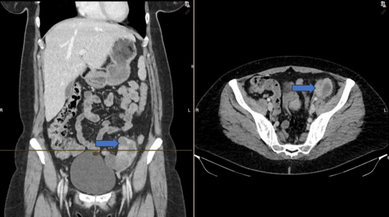
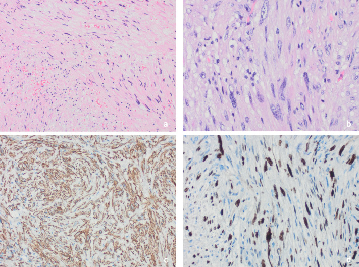
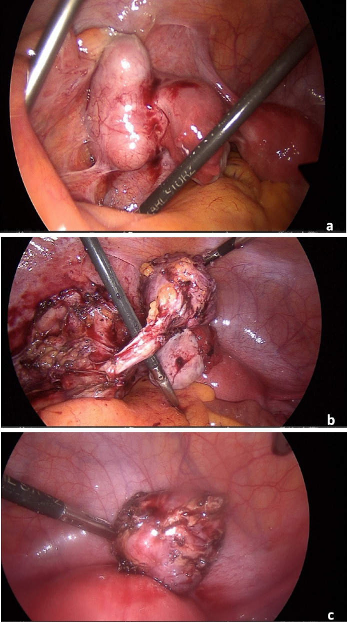
Leiomyosarcomas (LMS) arise from smooth muscle and represents only 6% of all sarcomas. LMS originating from major blood vessels, called vascular LMS, are detected mostly in the inferior vena cava. Arterial LMS are a rarity. We present a 43-year-old patient with a LMS arising from the left external iliac artery. The patient was referred to us with symptoms of left lower abdominal pain extending to the left limb and underwent a contrast computed tomography which suggested a suspicious mass near the left iliac vessels. She underwent laparoscopic excision of the tumour, whose histological examination revealed an LMS G2 arising from the external iliac artery. Immunohistochemically CD34, p53, Desmin, as well as smooth muscle actin, tested positive.

 求助内容:
求助内容: 应助结果提醒方式:


