{"title":"区域性毒性损伤后海马器官型切片模型小胶质细胞5种中间结构变化的评价。","authors":"A Jesus Trejos, A X Francis Schanne","doi":"10.1007/s10571-025-01545-1","DOIUrl":null,"url":null,"abstract":"<p><p>The dendritic cell of the CNS, the microglia (MG), is an initiation point of the immunological response within the post-blood-brain barrier (BBB) compartment. Microglia drastically changes in response to cell stress to a much different non-dendritic morphologies. This investigation postulates that if the first MG responses to toxic injury are isolated and studied in greater morphological detail, there is much to be learned about microglia's metamorphosis from and M2 to an M1 state. The organotypic hippocampal slice was the experimental setting used to investigate microglial response to toxic injury; this isolates dendritic cell to post-BBB cells dynamics from the impact of nonspecific of in vivo blood-derived signaling. Within the context of biochemically verified precise toxic cell injury/death (induced with mercury or cyanide in combination with 2-deoxy-glucose) to a specific region within the hippocampal slice, MG's morphological response was evaluated. There was up to 35% increase in microglia activation proximally to injury (CA3 region) and no changes distally (DG region) when compared to control slices treated with PBS. Maximum microglia activation consisted of a 3 plus-fold increase in the distance between the nucleus membrane and the cell membrane, which underscores an extensive and quantifiable amount of membrane rearrangement. This quantification can be applied to contemporaneous AI image analysis algorithms to demarcate and quantify relative MG activation in and around a site of injury. In between baseline and activated MG morphologies, 5 intermediate morphologies (or structural variations) are described as it relates to its cell body, nucleus, and dendrites. The result from this study reconciles details of MG's structure to its holistic characteristics in relation to parenchymal cell stress.</p>","PeriodicalId":9742,"journal":{"name":"Cellular and Molecular Neurobiology","volume":"45 1","pages":"34"},"PeriodicalIF":4.8000,"publicationDate":"2025-04-09","publicationTypes":"Journal Article","fieldsOfStudy":null,"isOpenAccess":false,"openAccessPdf":"https://www.ncbi.nlm.nih.gov/pmc/articles/PMC11981971/pdf/","citationCount":"0","resultStr":"{\"title\":\"Evaluation of 5 Intermediate Structural Variations of Microglia Within an Organotypic Hippocampal Slice Model After Regionalized Toxic Injury.\",\"authors\":\"A Jesus Trejos, A X Francis Schanne\",\"doi\":\"10.1007/s10571-025-01545-1\",\"DOIUrl\":null,\"url\":null,\"abstract\":\"<p><p>The dendritic cell of the CNS, the microglia (MG), is an initiation point of the immunological response within the post-blood-brain barrier (BBB) compartment. Microglia drastically changes in response to cell stress to a much different non-dendritic morphologies. This investigation postulates that if the first MG responses to toxic injury are isolated and studied in greater morphological detail, there is much to be learned about microglia's metamorphosis from and M2 to an M1 state. The organotypic hippocampal slice was the experimental setting used to investigate microglial response to toxic injury; this isolates dendritic cell to post-BBB cells dynamics from the impact of nonspecific of in vivo blood-derived signaling. Within the context of biochemically verified precise toxic cell injury/death (induced with mercury or cyanide in combination with 2-deoxy-glucose) to a specific region within the hippocampal slice, MG's morphological response was evaluated. There was up to 35% increase in microglia activation proximally to injury (CA3 region) and no changes distally (DG region) when compared to control slices treated with PBS. Maximum microglia activation consisted of a 3 plus-fold increase in the distance between the nucleus membrane and the cell membrane, which underscores an extensive and quantifiable amount of membrane rearrangement. This quantification can be applied to contemporaneous AI image analysis algorithms to demarcate and quantify relative MG activation in and around a site of injury. In between baseline and activated MG morphologies, 5 intermediate morphologies (or structural variations) are described as it relates to its cell body, nucleus, and dendrites. The result from this study reconciles details of MG's structure to its holistic characteristics in relation to parenchymal cell stress.</p>\",\"PeriodicalId\":9742,\"journal\":{\"name\":\"Cellular and Molecular Neurobiology\",\"volume\":\"45 1\",\"pages\":\"34\"},\"PeriodicalIF\":4.8000,\"publicationDate\":\"2025-04-09\",\"publicationTypes\":\"Journal Article\",\"fieldsOfStudy\":null,\"isOpenAccess\":false,\"openAccessPdf\":\"https://www.ncbi.nlm.nih.gov/pmc/articles/PMC11981971/pdf/\",\"citationCount\":\"0\",\"resultStr\":null,\"platform\":\"Semanticscholar\",\"paperid\":null,\"PeriodicalName\":\"Cellular and Molecular Neurobiology\",\"FirstCategoryId\":\"3\",\"ListUrlMain\":\"https://doi.org/10.1007/s10571-025-01545-1\",\"RegionNum\":4,\"RegionCategory\":\"医学\",\"ArticlePicture\":[],\"TitleCN\":null,\"AbstractTextCN\":null,\"PMCID\":null,\"EPubDate\":\"\",\"PubModel\":\"\",\"JCR\":\"Q3\",\"JCRName\":\"CELL BIOLOGY\",\"Score\":null,\"Total\":0}","platform":"Semanticscholar","paperid":null,"PeriodicalName":"Cellular and Molecular Neurobiology","FirstCategoryId":"3","ListUrlMain":"https://doi.org/10.1007/s10571-025-01545-1","RegionNum":4,"RegionCategory":"医学","ArticlePicture":[],"TitleCN":null,"AbstractTextCN":null,"PMCID":null,"EPubDate":"","PubModel":"","JCR":"Q3","JCRName":"CELL BIOLOGY","Score":null,"Total":0}
Evaluation of 5 Intermediate Structural Variations of Microglia Within an Organotypic Hippocampal Slice Model After Regionalized Toxic Injury.
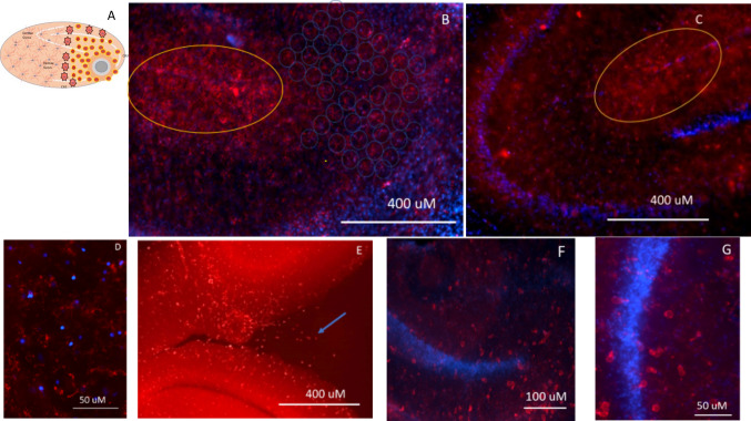
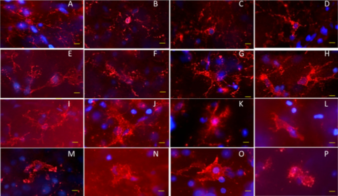
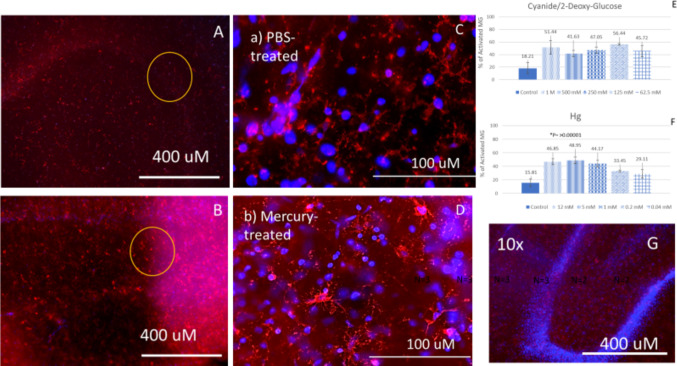
The dendritic cell of the CNS, the microglia (MG), is an initiation point of the immunological response within the post-blood-brain barrier (BBB) compartment. Microglia drastically changes in response to cell stress to a much different non-dendritic morphologies. This investigation postulates that if the first MG responses to toxic injury are isolated and studied in greater morphological detail, there is much to be learned about microglia's metamorphosis from and M2 to an M1 state. The organotypic hippocampal slice was the experimental setting used to investigate microglial response to toxic injury; this isolates dendritic cell to post-BBB cells dynamics from the impact of nonspecific of in vivo blood-derived signaling. Within the context of biochemically verified precise toxic cell injury/death (induced with mercury or cyanide in combination with 2-deoxy-glucose) to a specific region within the hippocampal slice, MG's morphological response was evaluated. There was up to 35% increase in microglia activation proximally to injury (CA3 region) and no changes distally (DG region) when compared to control slices treated with PBS. Maximum microglia activation consisted of a 3 plus-fold increase in the distance between the nucleus membrane and the cell membrane, which underscores an extensive and quantifiable amount of membrane rearrangement. This quantification can be applied to contemporaneous AI image analysis algorithms to demarcate and quantify relative MG activation in and around a site of injury. In between baseline and activated MG morphologies, 5 intermediate morphologies (or structural variations) are described as it relates to its cell body, nucleus, and dendrites. The result from this study reconciles details of MG's structure to its holistic characteristics in relation to parenchymal cell stress.
期刊介绍:
Cellular and Molecular Neurobiology publishes original research concerned with the analysis of neuronal and brain function at the cellular and subcellular levels. The journal offers timely, peer-reviewed articles that describe anatomic, genetic, physiologic, pharmacologic, and biochemical approaches to the study of neuronal function and the analysis of elementary mechanisms. Studies are presented on isolated mammalian tissues and intact animals, with investigations aimed at the molecular mechanisms or neuronal responses at the level of single cells. Cellular and Molecular Neurobiology also presents studies of the effects of neurons on other organ systems, such as analysis of the electrical or biochemical response to neurotransmitters or neurohormones on smooth muscle or gland cells.
 求助内容:
求助内容: 应助结果提醒方式:


