Natalie Smith, Helen McGuire, Barbara Fazekas de St Groth
{"title":"实现荧光团偶联单克隆抗体饱和的工作流程,用于生物标志物表达的稳健比较。","authors":"Natalie Smith, Helen McGuire, Barbara Fazekas de St Groth","doi":"10.1002/cyto.a.24938","DOIUrl":null,"url":null,"abstract":"<p>Antibody titration is an important step in every cytometric workflow, with the goal being to determine antibody concentrations that ensure highly reproducible results. When aiming to compare antigen expression between samples using mean or median fluorescence intensity (MFI), reagents should be used at a saturating concentration so that unavoidable variations in staining conditions do not affect the fluorescence signal. The recommended concentrations of commercially available fluorophore-labeled monoclonal antibodies (mAbs) may not achieve plateau staining, and their saturating concentration may be too high to be experimentally useful. To address these common concerns, we present a novel method to achieve saturation of fluorophore-conjugated mAbs, by ‘spiking-in’ unlabelled antibody of the same clone. Here, we demonstrate the application of this workflow to human anti-CD3 (clone OKT3, mouse IgG2a) and anti-TCRαβ (clone IP26, mouse IgG1), two mAbs that do not achieve saturation at 2-fold above their commercially recommended concentrations. First, the saturating concentration of unlabelled (purified) OKT3 and IP26 was determined by detection with a fluorophore-labeled anti-mouse IgG (H + L) secondary antibody. Titration curves of unlabelled and labeled mAbs were compared for each clone to determine whether labeling had resulted in any loss in binding activity. Unlabelled antibody was then ‘spiked’ into the labeled antibody at varying ratios, and those that achieved saturation while maintaining an adequate fluorescence signal were identified. We demonstrate that antibody saturation can be achieved with an optimized mixture of labeled and unlabelled antibody, while maintaining a clear signal from the fluorophore. While this workflow has only been applied to OKT3 and IP26, it has potential applicability for any antibody clone for which both labeled and unlabelled preparations are available. This method has significance for robust comparison of biomarker expression when fluorophore labeled reagents do not reach saturation under standard staining conditions.</p>","PeriodicalId":11068,"journal":{"name":"Cytometry Part A","volume":"107 6","pages":"378-389"},"PeriodicalIF":2.1000,"publicationDate":"2025-04-29","publicationTypes":"Journal Article","fieldsOfStudy":null,"isOpenAccess":false,"openAccessPdf":"https://onlinelibrary.wiley.com/doi/epdf/10.1002/cyto.a.24938","citationCount":"0","resultStr":"{\"title\":\"A Workflow to Achieve Saturation of Fluorophore-Conjugated Monoclonal Antibodies for Robust Comparison of Biomarker Expression\",\"authors\":\"Natalie Smith, Helen McGuire, Barbara Fazekas de St Groth\",\"doi\":\"10.1002/cyto.a.24938\",\"DOIUrl\":null,\"url\":null,\"abstract\":\"<p>Antibody titration is an important step in every cytometric workflow, with the goal being to determine antibody concentrations that ensure highly reproducible results. When aiming to compare antigen expression between samples using mean or median fluorescence intensity (MFI), reagents should be used at a saturating concentration so that unavoidable variations in staining conditions do not affect the fluorescence signal. The recommended concentrations of commercially available fluorophore-labeled monoclonal antibodies (mAbs) may not achieve plateau staining, and their saturating concentration may be too high to be experimentally useful. To address these common concerns, we present a novel method to achieve saturation of fluorophore-conjugated mAbs, by ‘spiking-in’ unlabelled antibody of the same clone. Here, we demonstrate the application of this workflow to human anti-CD3 (clone OKT3, mouse IgG2a) and anti-TCRαβ (clone IP26, mouse IgG1), two mAbs that do not achieve saturation at 2-fold above their commercially recommended concentrations. First, the saturating concentration of unlabelled (purified) OKT3 and IP26 was determined by detection with a fluorophore-labeled anti-mouse IgG (H + L) secondary antibody. Titration curves of unlabelled and labeled mAbs were compared for each clone to determine whether labeling had resulted in any loss in binding activity. Unlabelled antibody was then ‘spiked’ into the labeled antibody at varying ratios, and those that achieved saturation while maintaining an adequate fluorescence signal were identified. We demonstrate that antibody saturation can be achieved with an optimized mixture of labeled and unlabelled antibody, while maintaining a clear signal from the fluorophore. While this workflow has only been applied to OKT3 and IP26, it has potential applicability for any antibody clone for which both labeled and unlabelled preparations are available. This method has significance for robust comparison of biomarker expression when fluorophore labeled reagents do not reach saturation under standard staining conditions.</p>\",\"PeriodicalId\":11068,\"journal\":{\"name\":\"Cytometry Part A\",\"volume\":\"107 6\",\"pages\":\"378-389\"},\"PeriodicalIF\":2.1000,\"publicationDate\":\"2025-04-29\",\"publicationTypes\":\"Journal Article\",\"fieldsOfStudy\":null,\"isOpenAccess\":false,\"openAccessPdf\":\"https://onlinelibrary.wiley.com/doi/epdf/10.1002/cyto.a.24938\",\"citationCount\":\"0\",\"resultStr\":null,\"platform\":\"Semanticscholar\",\"paperid\":null,\"PeriodicalName\":\"Cytometry Part A\",\"FirstCategoryId\":\"99\",\"ListUrlMain\":\"https://onlinelibrary.wiley.com/doi/10.1002/cyto.a.24938\",\"RegionNum\":4,\"RegionCategory\":\"生物学\",\"ArticlePicture\":[],\"TitleCN\":null,\"AbstractTextCN\":null,\"PMCID\":null,\"EPubDate\":\"\",\"PubModel\":\"\",\"JCR\":\"Q3\",\"JCRName\":\"BIOCHEMICAL RESEARCH METHODS\",\"Score\":null,\"Total\":0}","platform":"Semanticscholar","paperid":null,"PeriodicalName":"Cytometry Part A","FirstCategoryId":"99","ListUrlMain":"https://onlinelibrary.wiley.com/doi/10.1002/cyto.a.24938","RegionNum":4,"RegionCategory":"生物学","ArticlePicture":[],"TitleCN":null,"AbstractTextCN":null,"PMCID":null,"EPubDate":"","PubModel":"","JCR":"Q3","JCRName":"BIOCHEMICAL RESEARCH METHODS","Score":null,"Total":0}
A Workflow to Achieve Saturation of Fluorophore-Conjugated Monoclonal Antibodies for Robust Comparison of Biomarker Expression
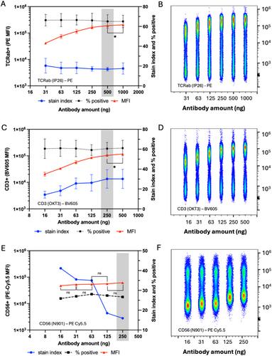
Antibody titration is an important step in every cytometric workflow, with the goal being to determine antibody concentrations that ensure highly reproducible results. When aiming to compare antigen expression between samples using mean or median fluorescence intensity (MFI), reagents should be used at a saturating concentration so that unavoidable variations in staining conditions do not affect the fluorescence signal. The recommended concentrations of commercially available fluorophore-labeled monoclonal antibodies (mAbs) may not achieve plateau staining, and their saturating concentration may be too high to be experimentally useful. To address these common concerns, we present a novel method to achieve saturation of fluorophore-conjugated mAbs, by ‘spiking-in’ unlabelled antibody of the same clone. Here, we demonstrate the application of this workflow to human anti-CD3 (clone OKT3, mouse IgG2a) and anti-TCRαβ (clone IP26, mouse IgG1), two mAbs that do not achieve saturation at 2-fold above their commercially recommended concentrations. First, the saturating concentration of unlabelled (purified) OKT3 and IP26 was determined by detection with a fluorophore-labeled anti-mouse IgG (H + L) secondary antibody. Titration curves of unlabelled and labeled mAbs were compared for each clone to determine whether labeling had resulted in any loss in binding activity. Unlabelled antibody was then ‘spiked’ into the labeled antibody at varying ratios, and those that achieved saturation while maintaining an adequate fluorescence signal were identified. We demonstrate that antibody saturation can be achieved with an optimized mixture of labeled and unlabelled antibody, while maintaining a clear signal from the fluorophore. While this workflow has only been applied to OKT3 and IP26, it has potential applicability for any antibody clone for which both labeled and unlabelled preparations are available. This method has significance for robust comparison of biomarker expression when fluorophore labeled reagents do not reach saturation under standard staining conditions.
期刊介绍:
Cytometry Part A, the journal of quantitative single-cell analysis, features original research reports and reviews of innovative scientific studies employing quantitative single-cell measurement, separation, manipulation, and modeling techniques, as well as original articles on mechanisms of molecular and cellular functions obtained by cytometry techniques.
The journal welcomes submissions from multiple research fields that fully embrace the study of the cytome:
Biomedical Instrumentation Engineering
Biophotonics
Bioinformatics
Cell Biology
Computational Biology
Data Science
Immunology
Parasitology
Microbiology
Neuroscience
Cancer
Stem Cells
Tissue Regeneration.
 求助内容:
求助内容: 应助结果提醒方式:


