Chi-Chang Weng, Chao-Chih Chiang, Yi-Hsiu Chung, Yi-Pei Ho, Yu-Chuan Chang, Ing-Tsung Hsiao, Robert H Mach
{"title":"乳腺癌动物模型中PARP抑制剂的简化放射性标记方法的评价。","authors":"Chi-Chang Weng, Chao-Chih Chiang, Yi-Hsiu Chung, Yi-Pei Ho, Yu-Chuan Chang, Ing-Tsung Hsiao, Robert H Mach","doi":"10.1186/s13550-025-01236-4","DOIUrl":null,"url":null,"abstract":"<p><strong>Background: </strong>Several poly (adenosine diphosphate-ribose) polymerase (PARP) inhibitors were recently approved by the US Food and Drug Administration for use in cancer treatment. To facilitate the discovery of novel PARP-targeting ligands, a radioiodinated ligand, I-125-KX1, was developed and validated for its specificity to PARP-1; however, its preparation procedure is time-consuming. The present study employed a solid-phase extraction (SPE) method in the radiolabeling procedure of I-123/125-KX1 and evaluated its binding specificity by using receptor binding assays, autoradiography, and in vivo single photon emission computed tomography (SPECT) imaging technique.</p><p><strong>Results: </strong>Through the incorporation of the SPE purification method as the final step in the radioiodination procedure, the resultant product I-123/125-KX1 exhibited high radiochemical purity (> 99%) and an acceptable radiochemical yield (58.6% for I-123-KX1, 73.3% for I-125-KX1). The binding characteristics of this radiotracer were validated through saturation binding assays conducted on MDA-MB-231 and MCF-7 cells. The K<sub>d</sub> values obtained for the tracer (~ 1.0 nM) was consistent with values reported in the literature, and the B<sub>max</sub> values of these two cell lines (2017 ± 178 fmol/mg on MDA-MB-231 vs. 1393 ± 105 fmol/mg on MCF-7) were in line with the results from Western blot analyses. To demonstrate the in vivo imaging ability of I-123-KX1 prepared in this study, an MDA-MB-231 tumor animal model was used and the tracer displayed a suitable uptake on the tumor tissues (6.9 ± 0.8%ID/mL). The binding specificity of the SPE-purified I-125-KX1 was further verified using in vitro autoradiography in conjunction with various PARP inhibitors. Additionally, an anti-PARP-1 immunohistochemistry experiment was conducted, which revealed that the autoradiograms of the radiotracer displayed a similar pattern.</p><p><strong>Conclusions: </strong>This suggests that the I-123/125-KX1 prepared using the SPE method showed some comparable properties to those from the traditional method, indicating its potential suitability for future radioligand preparation in PARP studies. However, further characterization studies may be needed to confirm its efficacy.</p>","PeriodicalId":11611,"journal":{"name":"EJNMMI Research","volume":"15 1","pages":"50"},"PeriodicalIF":3.1000,"publicationDate":"2025-04-29","publicationTypes":"Journal Article","fieldsOfStudy":null,"isOpenAccess":false,"openAccessPdf":"https://www.ncbi.nlm.nih.gov/pmc/articles/PMC12040802/pdf/","citationCount":"0","resultStr":"{\"title\":\"Evaluation of a simplified radiolabeling method for a PARP inhibitor in an animal model of breast cancer.\",\"authors\":\"Chi-Chang Weng, Chao-Chih Chiang, Yi-Hsiu Chung, Yi-Pei Ho, Yu-Chuan Chang, Ing-Tsung Hsiao, Robert H Mach\",\"doi\":\"10.1186/s13550-025-01236-4\",\"DOIUrl\":null,\"url\":null,\"abstract\":\"<p><strong>Background: </strong>Several poly (adenosine diphosphate-ribose) polymerase (PARP) inhibitors were recently approved by the US Food and Drug Administration for use in cancer treatment. To facilitate the discovery of novel PARP-targeting ligands, a radioiodinated ligand, I-125-KX1, was developed and validated for its specificity to PARP-1; however, its preparation procedure is time-consuming. The present study employed a solid-phase extraction (SPE) method in the radiolabeling procedure of I-123/125-KX1 and evaluated its binding specificity by using receptor binding assays, autoradiography, and in vivo single photon emission computed tomography (SPECT) imaging technique.</p><p><strong>Results: </strong>Through the incorporation of the SPE purification method as the final step in the radioiodination procedure, the resultant product I-123/125-KX1 exhibited high radiochemical purity (> 99%) and an acceptable radiochemical yield (58.6% for I-123-KX1, 73.3% for I-125-KX1). The binding characteristics of this radiotracer were validated through saturation binding assays conducted on MDA-MB-231 and MCF-7 cells. The K<sub>d</sub> values obtained for the tracer (~ 1.0 nM) was consistent with values reported in the literature, and the B<sub>max</sub> values of these two cell lines (2017 ± 178 fmol/mg on MDA-MB-231 vs. 1393 ± 105 fmol/mg on MCF-7) were in line with the results from Western blot analyses. To demonstrate the in vivo imaging ability of I-123-KX1 prepared in this study, an MDA-MB-231 tumor animal model was used and the tracer displayed a suitable uptake on the tumor tissues (6.9 ± 0.8%ID/mL). The binding specificity of the SPE-purified I-125-KX1 was further verified using in vitro autoradiography in conjunction with various PARP inhibitors. Additionally, an anti-PARP-1 immunohistochemistry experiment was conducted, which revealed that the autoradiograms of the radiotracer displayed a similar pattern.</p><p><strong>Conclusions: </strong>This suggests that the I-123/125-KX1 prepared using the SPE method showed some comparable properties to those from the traditional method, indicating its potential suitability for future radioligand preparation in PARP studies. However, further characterization studies may be needed to confirm its efficacy.</p>\",\"PeriodicalId\":11611,\"journal\":{\"name\":\"EJNMMI Research\",\"volume\":\"15 1\",\"pages\":\"50\"},\"PeriodicalIF\":3.1000,\"publicationDate\":\"2025-04-29\",\"publicationTypes\":\"Journal Article\",\"fieldsOfStudy\":null,\"isOpenAccess\":false,\"openAccessPdf\":\"https://www.ncbi.nlm.nih.gov/pmc/articles/PMC12040802/pdf/\",\"citationCount\":\"0\",\"resultStr\":null,\"platform\":\"Semanticscholar\",\"paperid\":null,\"PeriodicalName\":\"EJNMMI Research\",\"FirstCategoryId\":\"3\",\"ListUrlMain\":\"https://doi.org/10.1186/s13550-025-01236-4\",\"RegionNum\":3,\"RegionCategory\":\"医学\",\"ArticlePicture\":[],\"TitleCN\":null,\"AbstractTextCN\":null,\"PMCID\":null,\"EPubDate\":\"\",\"PubModel\":\"\",\"JCR\":\"Q1\",\"JCRName\":\"RADIOLOGY, NUCLEAR MEDICINE & MEDICAL IMAGING\",\"Score\":null,\"Total\":0}","platform":"Semanticscholar","paperid":null,"PeriodicalName":"EJNMMI Research","FirstCategoryId":"3","ListUrlMain":"https://doi.org/10.1186/s13550-025-01236-4","RegionNum":3,"RegionCategory":"医学","ArticlePicture":[],"TitleCN":null,"AbstractTextCN":null,"PMCID":null,"EPubDate":"","PubModel":"","JCR":"Q1","JCRName":"RADIOLOGY, NUCLEAR MEDICINE & MEDICAL IMAGING","Score":null,"Total":0}
Evaluation of a simplified radiolabeling method for a PARP inhibitor in an animal model of breast cancer.
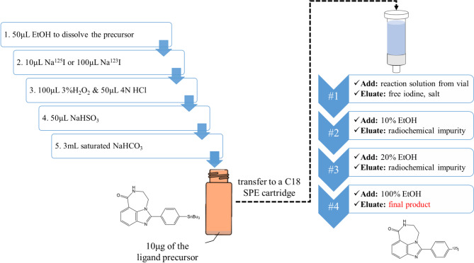
Background: Several poly (adenosine diphosphate-ribose) polymerase (PARP) inhibitors were recently approved by the US Food and Drug Administration for use in cancer treatment. To facilitate the discovery of novel PARP-targeting ligands, a radioiodinated ligand, I-125-KX1, was developed and validated for its specificity to PARP-1; however, its preparation procedure is time-consuming. The present study employed a solid-phase extraction (SPE) method in the radiolabeling procedure of I-123/125-KX1 and evaluated its binding specificity by using receptor binding assays, autoradiography, and in vivo single photon emission computed tomography (SPECT) imaging technique.
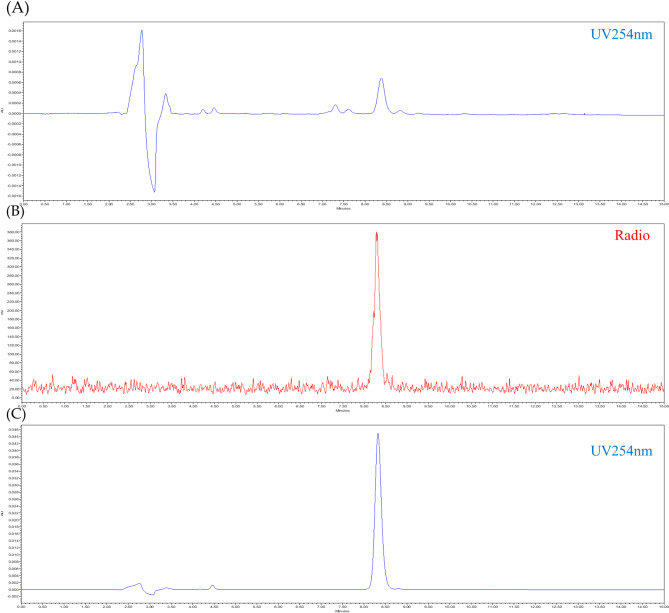
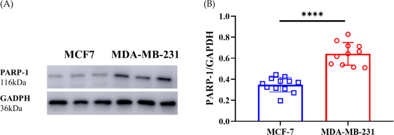
Results: Through the incorporation of the SPE purification method as the final step in the radioiodination procedure, the resultant product I-123/125-KX1 exhibited high radiochemical purity (> 99%) and an acceptable radiochemical yield (58.6% for I-123-KX1, 73.3% for I-125-KX1). The binding characteristics of this radiotracer were validated through saturation binding assays conducted on MDA-MB-231 and MCF-7 cells. The Kd values obtained for the tracer (~ 1.0 nM) was consistent with values reported in the literature, and the Bmax values of these two cell lines (2017 ± 178 fmol/mg on MDA-MB-231 vs. 1393 ± 105 fmol/mg on MCF-7) were in line with the results from Western blot analyses. To demonstrate the in vivo imaging ability of I-123-KX1 prepared in this study, an MDA-MB-231 tumor animal model was used and the tracer displayed a suitable uptake on the tumor tissues (6.9 ± 0.8%ID/mL). The binding specificity of the SPE-purified I-125-KX1 was further verified using in vitro autoradiography in conjunction with various PARP inhibitors. Additionally, an anti-PARP-1 immunohistochemistry experiment was conducted, which revealed that the autoradiograms of the radiotracer displayed a similar pattern.
Conclusions: This suggests that the I-123/125-KX1 prepared using the SPE method showed some comparable properties to those from the traditional method, indicating its potential suitability for future radioligand preparation in PARP studies. However, further characterization studies may be needed to confirm its efficacy.
EJNMMI ResearchRADIOLOGY, NUCLEAR MEDICINE & MEDICAL IMAGING&nb-
CiteScore
5.90
自引率
3.10%
发文量
72
审稿时长
13 weeks
期刊介绍:
EJNMMI Research publishes new basic, translational and clinical research in the field of nuclear medicine and molecular imaging. Regular features include original research articles, rapid communication of preliminary data on innovative research, interesting case reports, editorials, and letters to the editor. Educational articles on basic sciences, fundamental aspects and controversy related to pre-clinical and clinical research or ethical aspects of research are also welcome. Timely reviews provide updates on current applications, issues in imaging research and translational aspects of nuclear medicine and molecular imaging technologies.
The main emphasis is placed on the development of targeted imaging with radiopharmaceuticals within the broader context of molecular probes to enhance understanding and characterisation of the complex biological processes underlying disease and to develop, test and guide new treatment modalities, including radionuclide therapy.

 求助内容:
求助内容: 应助结果提醒方式:


