Gaojun Lu, Peilong Zhang, Sara Ricciardi, Ruotian Wang, Chen Wang, Kun Qian, Giuseppe Cardillo, Yi Zhang
{"title":"COVID-19大流行期间胸部计算机断层扫描发现偶发纵隔肿块。","authors":"Gaojun Lu, Peilong Zhang, Sara Ricciardi, Ruotian Wang, Chen Wang, Kun Qian, Giuseppe Cardillo, Yi Zhang","doi":"10.1093/ejcts/ezaf140","DOIUrl":null,"url":null,"abstract":"<p><strong>Objectives: </strong>The prevalence of mediastinal masses in large-scale populations in China has been rarely reported. During COVID19 pandemic, many incidentalomas were reported due to the large amount of chest computed tomography scan performed in emergency setting.</p><p><strong>Methods: </strong>Retrospective analysis of emergency chest computed tomography scans (February 2020-February 2021) for COVID-19 screening, including mediastinal abnormalities (excluding lymph nodes, dysplasia, pneumomediastinum and other non-mass alterations), with computed tomography features, diagnostic workup and 1 year follow-up data were reviewed.</p><p><strong>Results: </strong>Of the 40 112 patients [mean age 54.5 (17.2) years; male-to-female ratio 1.02:1] screened for COVID-19, 293 (0.73%) had mediastinal masses of which 223 (0.56%) located in the anterior mediastinum. As participants aged, the prevalence tended to increase (P < 0.001). The prevalence was not different between the sexes (P = 0.635). An oval shape, anterior mediastinal location, and thymus involvement were the most common computed tomography characteristics. Surgery confirmed 11.3% (33 of 293) of nodal lesions, with a benign to malignant ratio of 51.4: 48.5. A computed tomography scan follow-up was conducted in 32.3% (84/260) of the patients, and in 82.1% (69/84) of cases the lesion was stable. Additionally, mediastinal masses were detected in 7.7% (20/260) of elderly patients who passed away soon after their primary disease worsened.</p><p><strong>Conclusions: </strong>In Chinese COVID-19 screening chest computed tomography, the prevalence of all mediastinal masses and anterior mediastinal masses was 0.73% and 0.56%, respectively. Findings support risk-stratified management: growing/suspicious lesions warrant intervention versus surveillance for stable masses. Standardized protocols and multidisciplinary consensus are critical.</p>","PeriodicalId":11938,"journal":{"name":"European Journal of Cardio-Thoracic Surgery","volume":"67 4","pages":""},"PeriodicalIF":3.0000,"publicationDate":"2025-03-28","publicationTypes":"Journal Article","fieldsOfStudy":null,"isOpenAccess":false,"openAccessPdf":"https://www.ncbi.nlm.nih.gov/pmc/articles/PMC12043007/pdf/","citationCount":"0","resultStr":"{\"title\":\"Incidental mediastinal masses detected on chest computed tomography scans during the COVID-19 pandemic.\",\"authors\":\"Gaojun Lu, Peilong Zhang, Sara Ricciardi, Ruotian Wang, Chen Wang, Kun Qian, Giuseppe Cardillo, Yi Zhang\",\"doi\":\"10.1093/ejcts/ezaf140\",\"DOIUrl\":null,\"url\":null,\"abstract\":\"<p><strong>Objectives: </strong>The prevalence of mediastinal masses in large-scale populations in China has been rarely reported. During COVID19 pandemic, many incidentalomas were reported due to the large amount of chest computed tomography scan performed in emergency setting.</p><p><strong>Methods: </strong>Retrospective analysis of emergency chest computed tomography scans (February 2020-February 2021) for COVID-19 screening, including mediastinal abnormalities (excluding lymph nodes, dysplasia, pneumomediastinum and other non-mass alterations), with computed tomography features, diagnostic workup and 1 year follow-up data were reviewed.</p><p><strong>Results: </strong>Of the 40 112 patients [mean age 54.5 (17.2) years; male-to-female ratio 1.02:1] screened for COVID-19, 293 (0.73%) had mediastinal masses of which 223 (0.56%) located in the anterior mediastinum. As participants aged, the prevalence tended to increase (P < 0.001). The prevalence was not different between the sexes (P = 0.635). An oval shape, anterior mediastinal location, and thymus involvement were the most common computed tomography characteristics. Surgery confirmed 11.3% (33 of 293) of nodal lesions, with a benign to malignant ratio of 51.4: 48.5. A computed tomography scan follow-up was conducted in 32.3% (84/260) of the patients, and in 82.1% (69/84) of cases the lesion was stable. Additionally, mediastinal masses were detected in 7.7% (20/260) of elderly patients who passed away soon after their primary disease worsened.</p><p><strong>Conclusions: </strong>In Chinese COVID-19 screening chest computed tomography, the prevalence of all mediastinal masses and anterior mediastinal masses was 0.73% and 0.56%, respectively. Findings support risk-stratified management: growing/suspicious lesions warrant intervention versus surveillance for stable masses. Standardized protocols and multidisciplinary consensus are critical.</p>\",\"PeriodicalId\":11938,\"journal\":{\"name\":\"European Journal of Cardio-Thoracic Surgery\",\"volume\":\"67 4\",\"pages\":\"\"},\"PeriodicalIF\":3.0000,\"publicationDate\":\"2025-03-28\",\"publicationTypes\":\"Journal Article\",\"fieldsOfStudy\":null,\"isOpenAccess\":false,\"openAccessPdf\":\"https://www.ncbi.nlm.nih.gov/pmc/articles/PMC12043007/pdf/\",\"citationCount\":\"0\",\"resultStr\":null,\"platform\":\"Semanticscholar\",\"paperid\":null,\"PeriodicalName\":\"European Journal of Cardio-Thoracic Surgery\",\"FirstCategoryId\":\"3\",\"ListUrlMain\":\"https://doi.org/10.1093/ejcts/ezaf140\",\"RegionNum\":2,\"RegionCategory\":\"医学\",\"ArticlePicture\":[],\"TitleCN\":null,\"AbstractTextCN\":null,\"PMCID\":null,\"EPubDate\":\"\",\"PubModel\":\"\",\"JCR\":\"Q2\",\"JCRName\":\"CARDIAC & CARDIOVASCULAR SYSTEMS\",\"Score\":null,\"Total\":0}","platform":"Semanticscholar","paperid":null,"PeriodicalName":"European Journal of Cardio-Thoracic Surgery","FirstCategoryId":"3","ListUrlMain":"https://doi.org/10.1093/ejcts/ezaf140","RegionNum":2,"RegionCategory":"医学","ArticlePicture":[],"TitleCN":null,"AbstractTextCN":null,"PMCID":null,"EPubDate":"","PubModel":"","JCR":"Q2","JCRName":"CARDIAC & CARDIOVASCULAR SYSTEMS","Score":null,"Total":0}
Incidental mediastinal masses detected on chest computed tomography scans during the COVID-19 pandemic.
Objectives: The prevalence of mediastinal masses in large-scale populations in China has been rarely reported. During COVID19 pandemic, many incidentalomas were reported due to the large amount of chest computed tomography scan performed in emergency setting.
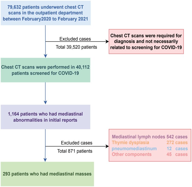
Methods: Retrospective analysis of emergency chest computed tomography scans (February 2020-February 2021) for COVID-19 screening, including mediastinal abnormalities (excluding lymph nodes, dysplasia, pneumomediastinum and other non-mass alterations), with computed tomography features, diagnostic workup and 1 year follow-up data were reviewed.
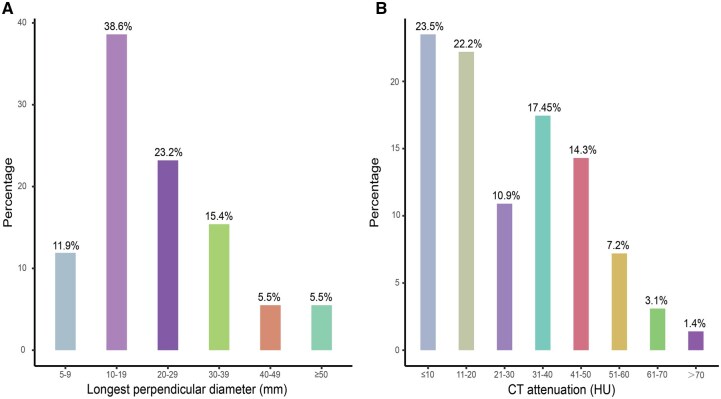
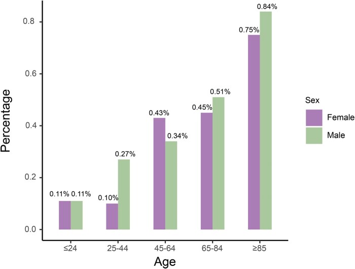
Results: Of the 40 112 patients [mean age 54.5 (17.2) years; male-to-female ratio 1.02:1] screened for COVID-19, 293 (0.73%) had mediastinal masses of which 223 (0.56%) located in the anterior mediastinum. As participants aged, the prevalence tended to increase (P < 0.001). The prevalence was not different between the sexes (P = 0.635). An oval shape, anterior mediastinal location, and thymus involvement were the most common computed tomography characteristics. Surgery confirmed 11.3% (33 of 293) of nodal lesions, with a benign to malignant ratio of 51.4: 48.5. A computed tomography scan follow-up was conducted in 32.3% (84/260) of the patients, and in 82.1% (69/84) of cases the lesion was stable. Additionally, mediastinal masses were detected in 7.7% (20/260) of elderly patients who passed away soon after their primary disease worsened.
Conclusions: In Chinese COVID-19 screening chest computed tomography, the prevalence of all mediastinal masses and anterior mediastinal masses was 0.73% and 0.56%, respectively. Findings support risk-stratified management: growing/suspicious lesions warrant intervention versus surveillance for stable masses. Standardized protocols and multidisciplinary consensus are critical.
期刊介绍:
The primary aim of the European Journal of Cardio-Thoracic Surgery is to provide a medium for the publication of high-quality original scientific reports documenting progress in cardiac and thoracic surgery. The journal publishes reports of significant clinical and experimental advances related to surgery of the heart, the great vessels and the chest. The European Journal of Cardio-Thoracic Surgery is an international journal and accepts submissions from all regions. The journal is supported by a number of leading European societies.
 求助内容:
求助内容: 应助结果提醒方式:


