{"title":"胎儿超声诊断脐动脉血栓形成。","authors":"Yushi Abe, Kazunori Ueno, Saki Tamura, Haruko Ariga, Jun Miyauchi, Hiroyuki Nakagawa","doi":"10.1515/crpm-2024-0017","DOIUrl":null,"url":null,"abstract":"<p><strong>Objectives: </strong>Umbilical artery thrombosis (UAT) is a rare and severe condition associated with grave perinatal outcomes, including intrauterine fetal death. This case report presents the case of a 38-year-old woman (gravida 3, para 1) of Japanese ethnicity, with a history of one spontaneous miscarriage, who conceived through micro-insemination and blastocyst transfer.</p><p><strong>Case presentation: </strong>Initial patient screening at 30 weeks and 6 days of gestation revealed normal fetal development, with two umbilical arteries and one umbilical vein. However, at 34 weeks and 5 days of gestation, we observed reduced fetal movements and the absence of accelerations on cardiotocography. Subsequent color Doppler examination revealed cessation of blood flow across a broad area in one umbilical artery and a strongly curved umbilical vein surrounding the blood flow of the other artery. These formed the 'orange grab sign,' suggestive of UAT. Evaluations of blood flow in other areas revealed unremarkable findings. We performed an emergency cesarean section owing to fetal distress. The mother and newborn were healthy and discharged as healthy. The 1-month check-up revealed no abnormalities in the child. Pathological examination of the umbilical cord revealed fibrin-based thrombus formation along the length of one artery, confirmed to be an umbilical artery.</p><p><strong>Conclusions: </strong>In the present case report, we presented the diagnostic challenges of UAT. Furthermore, we highlighted the need for timely intervention by comparing the number of umbilical vessels among previous ultrasound findings and verifying the presence of the 'orange grab sign.'</p>","PeriodicalId":9617,"journal":{"name":"Case Reports in Perinatal Medicine","volume":"13 1","pages":"20240017"},"PeriodicalIF":0.2000,"publicationDate":"2024-09-12","publicationTypes":"Journal Article","fieldsOfStudy":null,"isOpenAccess":false,"openAccessPdf":"https://www.ncbi.nlm.nih.gov/pmc/articles/PMC12048147/pdf/","citationCount":"0","resultStr":"{\"title\":\"Umbilical artery thrombosis diagnosed by fetal ultrasound.\",\"authors\":\"Yushi Abe, Kazunori Ueno, Saki Tamura, Haruko Ariga, Jun Miyauchi, Hiroyuki Nakagawa\",\"doi\":\"10.1515/crpm-2024-0017\",\"DOIUrl\":null,\"url\":null,\"abstract\":\"<p><strong>Objectives: </strong>Umbilical artery thrombosis (UAT) is a rare and severe condition associated with grave perinatal outcomes, including intrauterine fetal death. This case report presents the case of a 38-year-old woman (gravida 3, para 1) of Japanese ethnicity, with a history of one spontaneous miscarriage, who conceived through micro-insemination and blastocyst transfer.</p><p><strong>Case presentation: </strong>Initial patient screening at 30 weeks and 6 days of gestation revealed normal fetal development, with two umbilical arteries and one umbilical vein. However, at 34 weeks and 5 days of gestation, we observed reduced fetal movements and the absence of accelerations on cardiotocography. Subsequent color Doppler examination revealed cessation of blood flow across a broad area in one umbilical artery and a strongly curved umbilical vein surrounding the blood flow of the other artery. These formed the 'orange grab sign,' suggestive of UAT. Evaluations of blood flow in other areas revealed unremarkable findings. We performed an emergency cesarean section owing to fetal distress. The mother and newborn were healthy and discharged as healthy. The 1-month check-up revealed no abnormalities in the child. Pathological examination of the umbilical cord revealed fibrin-based thrombus formation along the length of one artery, confirmed to be an umbilical artery.</p><p><strong>Conclusions: </strong>In the present case report, we presented the diagnostic challenges of UAT. Furthermore, we highlighted the need for timely intervention by comparing the number of umbilical vessels among previous ultrasound findings and verifying the presence of the 'orange grab sign.'</p>\",\"PeriodicalId\":9617,\"journal\":{\"name\":\"Case Reports in Perinatal Medicine\",\"volume\":\"13 1\",\"pages\":\"20240017\"},\"PeriodicalIF\":0.2000,\"publicationDate\":\"2024-09-12\",\"publicationTypes\":\"Journal Article\",\"fieldsOfStudy\":null,\"isOpenAccess\":false,\"openAccessPdf\":\"https://www.ncbi.nlm.nih.gov/pmc/articles/PMC12048147/pdf/\",\"citationCount\":\"0\",\"resultStr\":null,\"platform\":\"Semanticscholar\",\"paperid\":null,\"PeriodicalName\":\"Case Reports in Perinatal Medicine\",\"FirstCategoryId\":\"1085\",\"ListUrlMain\":\"https://doi.org/10.1515/crpm-2024-0017\",\"RegionNum\":0,\"RegionCategory\":null,\"ArticlePicture\":[],\"TitleCN\":null,\"AbstractTextCN\":null,\"PMCID\":null,\"EPubDate\":\"2024/1/1 0:00:00\",\"PubModel\":\"eCollection\",\"JCR\":\"Q4\",\"JCRName\":\"OBSTETRICS & GYNECOLOGY\",\"Score\":null,\"Total\":0}","platform":"Semanticscholar","paperid":null,"PeriodicalName":"Case Reports in Perinatal Medicine","FirstCategoryId":"1085","ListUrlMain":"https://doi.org/10.1515/crpm-2024-0017","RegionNum":0,"RegionCategory":null,"ArticlePicture":[],"TitleCN":null,"AbstractTextCN":null,"PMCID":null,"EPubDate":"2024/1/1 0:00:00","PubModel":"eCollection","JCR":"Q4","JCRName":"OBSTETRICS & GYNECOLOGY","Score":null,"Total":0}
Umbilical artery thrombosis diagnosed by fetal ultrasound.
Objectives: Umbilical artery thrombosis (UAT) is a rare and severe condition associated with grave perinatal outcomes, including intrauterine fetal death. This case report presents the case of a 38-year-old woman (gravida 3, para 1) of Japanese ethnicity, with a history of one spontaneous miscarriage, who conceived through micro-insemination and blastocyst transfer.
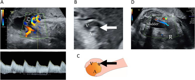
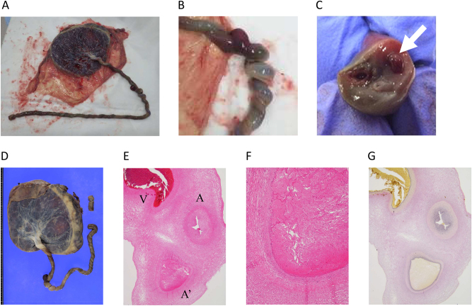
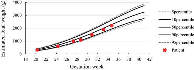
Case presentation: Initial patient screening at 30 weeks and 6 days of gestation revealed normal fetal development, with two umbilical arteries and one umbilical vein. However, at 34 weeks and 5 days of gestation, we observed reduced fetal movements and the absence of accelerations on cardiotocography. Subsequent color Doppler examination revealed cessation of blood flow across a broad area in one umbilical artery and a strongly curved umbilical vein surrounding the blood flow of the other artery. These formed the 'orange grab sign,' suggestive of UAT. Evaluations of blood flow in other areas revealed unremarkable findings. We performed an emergency cesarean section owing to fetal distress. The mother and newborn were healthy and discharged as healthy. The 1-month check-up revealed no abnormalities in the child. Pathological examination of the umbilical cord revealed fibrin-based thrombus formation along the length of one artery, confirmed to be an umbilical artery.
Conclusions: In the present case report, we presented the diagnostic challenges of UAT. Furthermore, we highlighted the need for timely intervention by comparing the number of umbilical vessels among previous ultrasound findings and verifying the presence of the 'orange grab sign.'
期刊介绍:
Case Reports in Perinatal Medicine is a double-blind peer-reviewed journal. The objective of the new journal is very similar to that of JPM. In addition to evidence-based studies, practitioners in clinical practice esteem especially exemplary reports of cases that reveal specific manifestations of diseases, its progress or its treatment. We consider case reports and series to be brief reports describing an isolated clinical case or a small number of cases. They may describe new or uncommon diagnoses, unusual outcomes or prognosis, new or infrequently used therapies and side effects of therapy not usually discovered in clinical trials. They represent the basic concept of experiences for studies on representative groups for further evidence-based research. The potential roles of case reports and case series are: Recognition and description of new diseases Detection of drug side effects (adverse or beneficial) Study of mechanisms of disease Medical education and audit Recognition of rare manifestations of disease.

 求助内容:
求助内容: 应助结果提醒方式:


