{"title":"Npc1基因突变损害小鼠肝细胞多系分化潜能。","authors":"Jichao Yang, Yuqiao Chang, Liang Qiao, Ganesh Dama, Yongli Lou, Juntang Lin","doi":"10.1007/s10565-025-10018-6","DOIUrl":null,"url":null,"abstract":"<p><strong>Objective: </strong>To investigate the effect of the Npc1 gene on the biological activity of Telocytes (TCs) in the liver and to provide theoretical support for further research on the biological activity of TCs.</p><p><strong>Methods: </strong>Primary liver tissue cultures (TCs) from neonatal Npc1<sup>+/+</sup> and Npc1<sup>-/-</sup> mice were extracted and cultured using an optimized type II collagenase-digestion protocol, and subsequently purified through a differential adhesion method. The growth state of TCs in both Npc1<sup>+/+</sup> and Npc1<sup>-/-</sup> groups was regularly observed under an inverted microscope, and the morphology of TCs under normal growth conditions was documented. The TCs were identified using scanning electron microscopy and immunofluorescence staining. To investigate the impact of the Npc1 gene on the multilineage differentiation potential of TCs, liver TCs from Npc1<sup>+/+</sup> and Npc1<sup>-/-</sup> groups were induced with adipogenic, osteogenic, and cardiomyoblastic differentiation solutions, respectively.</p><p><strong>Results: </strong>TCs cell surface markers such as co-expression of vimentin/CD34, vimentin/PDGF-α, and vimentin/c-Kit in Npc1<sup>+/+</sup> and Npc1<sup>-/-</sup> groups. \"Combined light and scanning electron microscopy revealed that the cellular structure of TCs from Npc1<sup>+/+</sup> and Npc1<sup>-/-</sup> groups was mainly composed of cell bodies and Telopodes (Tps). TCs exhibited small somata with fusiform, stellate, or spindle-shaped nuclei, depending on the number of Tps. The surface of TCs cell membrane was uneven, and there was no difference in morphology between the two groups. TCs had multilineage differentiation potential, and the positive rate of TCs induced in Npc1<sup>-/-</sup> group was significantly lower than that in the Npc1<sup>+/+</sup> group.</p><p><strong>Conclusion: </strong>Our findings demonstrate that NPC1 deficiency markedly attenuates hepatic TCs' multipotency of liver TCs to differentiate into adipocytes, osteoblasts, and cardiocytes, suggesting that NPC1 protein might affect the pluripotency of TCs by regulating the lipid transport pathway. This finding provides novel insights into TC-mediated mechanisms in NPC pathology and lays a theoretical foundation for regenerative medicine strategies targeting TCs.</p>","PeriodicalId":9672,"journal":{"name":"Cell Biology and Toxicology","volume":"41 1","pages":"71"},"PeriodicalIF":5.9000,"publicationDate":"2025-04-21","publicationTypes":"Journal Article","fieldsOfStudy":null,"isOpenAccess":false,"openAccessPdf":"https://www.ncbi.nlm.nih.gov/pmc/articles/PMC12011650/pdf/","citationCount":"0","resultStr":"{\"title\":\"Npc1 gene mutation impairs multilineage differentiation potential of hepatic telocytes in murine models.\",\"authors\":\"Jichao Yang, Yuqiao Chang, Liang Qiao, Ganesh Dama, Yongli Lou, Juntang Lin\",\"doi\":\"10.1007/s10565-025-10018-6\",\"DOIUrl\":null,\"url\":null,\"abstract\":\"<p><strong>Objective: </strong>To investigate the effect of the Npc1 gene on the biological activity of Telocytes (TCs) in the liver and to provide theoretical support for further research on the biological activity of TCs.</p><p><strong>Methods: </strong>Primary liver tissue cultures (TCs) from neonatal Npc1<sup>+/+</sup> and Npc1<sup>-/-</sup> mice were extracted and cultured using an optimized type II collagenase-digestion protocol, and subsequently purified through a differential adhesion method. The growth state of TCs in both Npc1<sup>+/+</sup> and Npc1<sup>-/-</sup> groups was regularly observed under an inverted microscope, and the morphology of TCs under normal growth conditions was documented. The TCs were identified using scanning electron microscopy and immunofluorescence staining. To investigate the impact of the Npc1 gene on the multilineage differentiation potential of TCs, liver TCs from Npc1<sup>+/+</sup> and Npc1<sup>-/-</sup> groups were induced with adipogenic, osteogenic, and cardiomyoblastic differentiation solutions, respectively.</p><p><strong>Results: </strong>TCs cell surface markers such as co-expression of vimentin/CD34, vimentin/PDGF-α, and vimentin/c-Kit in Npc1<sup>+/+</sup> and Npc1<sup>-/-</sup> groups. \\\"Combined light and scanning electron microscopy revealed that the cellular structure of TCs from Npc1<sup>+/+</sup> and Npc1<sup>-/-</sup> groups was mainly composed of cell bodies and Telopodes (Tps). TCs exhibited small somata with fusiform, stellate, or spindle-shaped nuclei, depending on the number of Tps. The surface of TCs cell membrane was uneven, and there was no difference in morphology between the two groups. TCs had multilineage differentiation potential, and the positive rate of TCs induced in Npc1<sup>-/-</sup> group was significantly lower than that in the Npc1<sup>+/+</sup> group.</p><p><strong>Conclusion: </strong>Our findings demonstrate that NPC1 deficiency markedly attenuates hepatic TCs' multipotency of liver TCs to differentiate into adipocytes, osteoblasts, and cardiocytes, suggesting that NPC1 protein might affect the pluripotency of TCs by regulating the lipid transport pathway. This finding provides novel insights into TC-mediated mechanisms in NPC pathology and lays a theoretical foundation for regenerative medicine strategies targeting TCs.</p>\",\"PeriodicalId\":9672,\"journal\":{\"name\":\"Cell Biology and Toxicology\",\"volume\":\"41 1\",\"pages\":\"71\"},\"PeriodicalIF\":5.9000,\"publicationDate\":\"2025-04-21\",\"publicationTypes\":\"Journal Article\",\"fieldsOfStudy\":null,\"isOpenAccess\":false,\"openAccessPdf\":\"https://www.ncbi.nlm.nih.gov/pmc/articles/PMC12011650/pdf/\",\"citationCount\":\"0\",\"resultStr\":null,\"platform\":\"Semanticscholar\",\"paperid\":null,\"PeriodicalName\":\"Cell Biology and Toxicology\",\"FirstCategoryId\":\"3\",\"ListUrlMain\":\"https://doi.org/10.1007/s10565-025-10018-6\",\"RegionNum\":2,\"RegionCategory\":\"医学\",\"ArticlePicture\":[],\"TitleCN\":null,\"AbstractTextCN\":null,\"PMCID\":null,\"EPubDate\":\"\",\"PubModel\":\"\",\"JCR\":\"Q2\",\"JCRName\":\"CELL BIOLOGY\",\"Score\":null,\"Total\":0}","platform":"Semanticscholar","paperid":null,"PeriodicalName":"Cell Biology and Toxicology","FirstCategoryId":"3","ListUrlMain":"https://doi.org/10.1007/s10565-025-10018-6","RegionNum":2,"RegionCategory":"医学","ArticlePicture":[],"TitleCN":null,"AbstractTextCN":null,"PMCID":null,"EPubDate":"","PubModel":"","JCR":"Q2","JCRName":"CELL BIOLOGY","Score":null,"Total":0}
Npc1 gene mutation impairs multilineage differentiation potential of hepatic telocytes in murine models.
Objective: To investigate the effect of the Npc1 gene on the biological activity of Telocytes (TCs) in the liver and to provide theoretical support for further research on the biological activity of TCs.
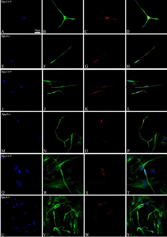
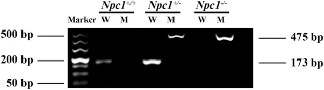
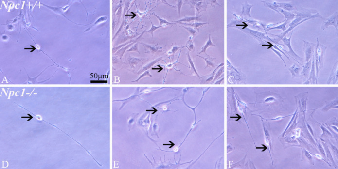
Methods: Primary liver tissue cultures (TCs) from neonatal Npc1+/+ and Npc1-/- mice were extracted and cultured using an optimized type II collagenase-digestion protocol, and subsequently purified through a differential adhesion method. The growth state of TCs in both Npc1+/+ and Npc1-/- groups was regularly observed under an inverted microscope, and the morphology of TCs under normal growth conditions was documented. The TCs were identified using scanning electron microscopy and immunofluorescence staining. To investigate the impact of the Npc1 gene on the multilineage differentiation potential of TCs, liver TCs from Npc1+/+ and Npc1-/- groups were induced with adipogenic, osteogenic, and cardiomyoblastic differentiation solutions, respectively.
Results: TCs cell surface markers such as co-expression of vimentin/CD34, vimentin/PDGF-α, and vimentin/c-Kit in Npc1+/+ and Npc1-/- groups. "Combined light and scanning electron microscopy revealed that the cellular structure of TCs from Npc1+/+ and Npc1-/- groups was mainly composed of cell bodies and Telopodes (Tps). TCs exhibited small somata with fusiform, stellate, or spindle-shaped nuclei, depending on the number of Tps. The surface of TCs cell membrane was uneven, and there was no difference in morphology between the two groups. TCs had multilineage differentiation potential, and the positive rate of TCs induced in Npc1-/- group was significantly lower than that in the Npc1+/+ group.
Conclusion: Our findings demonstrate that NPC1 deficiency markedly attenuates hepatic TCs' multipotency of liver TCs to differentiate into adipocytes, osteoblasts, and cardiocytes, suggesting that NPC1 protein might affect the pluripotency of TCs by regulating the lipid transport pathway. This finding provides novel insights into TC-mediated mechanisms in NPC pathology and lays a theoretical foundation for regenerative medicine strategies targeting TCs.
期刊介绍:
Cell Biology and Toxicology (CBT) is an international journal focused on clinical and translational research with an emphasis on molecular and cell biology, genetic and epigenetic heterogeneity, drug discovery and development, and molecular pharmacology and toxicology. CBT has a disease-specific scope prioritizing publications on gene and protein-based regulation, intracellular signaling pathway dysfunction, cell type-specific function, and systems in biomedicine in drug discovery and development. CBT publishes original articles with outstanding, innovative and significant findings, important reviews on recent research advances and issues of high current interest, opinion articles of leading edge science, and rapid communication or reports, on molecular mechanisms and therapies in diseases.
 求助内容:
求助内容: 应助结果提醒方式:


