{"title":"转子间骨折内侧骨折线空间分布的定量分析。","authors":"Miaotian Tang, Hao Zhou, Gaoxiang Xu, Dake Tong, Cheng Xu, Jiantao Li","doi":"10.1155/abb/6959877","DOIUrl":null,"url":null,"abstract":"<p><p>The integrity of the medial wall of the proximal femur is crucial for maintaining mechanical homeostasis. However, the impact of intertrochanteric fractures on the medial wall and the optimal diagnostic methodology remain unclear. We retrospectively analyzed CT data from 205 patients with intertrochanteric fractures. The lowest point of the medial fracture line was marked, and a standard coordinate system was established to record its spatial position. The association between AO, Evans, or Tang classification and different types of medial wall disruption was analyzed using Spearman correlation. The lowest point of the fracture line was located in the first quadrant of the proximal medial wall in 20 patients, with spatial coordinates of (6.44 ± 5.47, 6.14 ± 2.71). In 21 patients, it was in the second quadrant, with coordinates of (-7.23 ± 5.86, 8.31 ± 6.59). In 122 patients, it was in the third quadrant, with coordinates of (-9.59 ± 4.32, -24.43 ± 15.79), and in 42 patients, it was in the fourth quadrant, with coordinates of (8.18 ± 4.56, -18.20 ± 12.92). The Tang classification showed a stronger correlation with fracture instability (<i>r</i> = 0.40, <i>p</i> < 0.001) compared to the AO (<i>r</i> = 0.32, <i>p</i> < 0.001) and Evans (<i>r</i> = 0.38, <i>p</i> < 0.001) classifications. The medial wall of the proximal femur is significantly compromised in intertrochanteric fractures, with varying mechanical stability depending on the fracture type. The Tang classification effectively differentiates these stability differences, providing valuable guidance for clinical practice.</p>","PeriodicalId":8029,"journal":{"name":"Applied Bionics and Biomechanics","volume":"2025 ","pages":"6959877"},"PeriodicalIF":0.6000,"publicationDate":"2025-04-10","publicationTypes":"Journal Article","fieldsOfStudy":null,"isOpenAccess":false,"openAccessPdf":"https://www.ncbi.nlm.nih.gov/pmc/articles/PMC12006714/pdf/","citationCount":"0","resultStr":"{\"title\":\"Quantitative Analysis of Spatial Distribution of Medial Fracture Lines in Intertrochanteric Fractures.\",\"authors\":\"Miaotian Tang, Hao Zhou, Gaoxiang Xu, Dake Tong, Cheng Xu, Jiantao Li\",\"doi\":\"10.1155/abb/6959877\",\"DOIUrl\":null,\"url\":null,\"abstract\":\"<p><p>The integrity of the medial wall of the proximal femur is crucial for maintaining mechanical homeostasis. However, the impact of intertrochanteric fractures on the medial wall and the optimal diagnostic methodology remain unclear. We retrospectively analyzed CT data from 205 patients with intertrochanteric fractures. The lowest point of the medial fracture line was marked, and a standard coordinate system was established to record its spatial position. The association between AO, Evans, or Tang classification and different types of medial wall disruption was analyzed using Spearman correlation. The lowest point of the fracture line was located in the first quadrant of the proximal medial wall in 20 patients, with spatial coordinates of (6.44 ± 5.47, 6.14 ± 2.71). In 21 patients, it was in the second quadrant, with coordinates of (-7.23 ± 5.86, 8.31 ± 6.59). In 122 patients, it was in the third quadrant, with coordinates of (-9.59 ± 4.32, -24.43 ± 15.79), and in 42 patients, it was in the fourth quadrant, with coordinates of (8.18 ± 4.56, -18.20 ± 12.92). The Tang classification showed a stronger correlation with fracture instability (<i>r</i> = 0.40, <i>p</i> < 0.001) compared to the AO (<i>r</i> = 0.32, <i>p</i> < 0.001) and Evans (<i>r</i> = 0.38, <i>p</i> < 0.001) classifications. The medial wall of the proximal femur is significantly compromised in intertrochanteric fractures, with varying mechanical stability depending on the fracture type. The Tang classification effectively differentiates these stability differences, providing valuable guidance for clinical practice.</p>\",\"PeriodicalId\":8029,\"journal\":{\"name\":\"Applied Bionics and Biomechanics\",\"volume\":\"2025 \",\"pages\":\"6959877\"},\"PeriodicalIF\":0.6000,\"publicationDate\":\"2025-04-10\",\"publicationTypes\":\"Journal Article\",\"fieldsOfStudy\":null,\"isOpenAccess\":false,\"openAccessPdf\":\"https://www.ncbi.nlm.nih.gov/pmc/articles/PMC12006714/pdf/\",\"citationCount\":\"0\",\"resultStr\":null,\"platform\":\"Semanticscholar\",\"paperid\":null,\"PeriodicalName\":\"Applied Bionics and Biomechanics\",\"FirstCategoryId\":\"94\",\"ListUrlMain\":\"https://doi.org/10.1155/abb/6959877\",\"RegionNum\":4,\"RegionCategory\":\"计算机科学\",\"ArticlePicture\":[],\"TitleCN\":null,\"AbstractTextCN\":null,\"PMCID\":null,\"EPubDate\":\"2025/1/1 0:00:00\",\"PubModel\":\"eCollection\",\"JCR\":\"Q3\",\"JCRName\":\"ENGINEERING, BIOMEDICAL\",\"Score\":null,\"Total\":0}","platform":"Semanticscholar","paperid":null,"PeriodicalName":"Applied Bionics and Biomechanics","FirstCategoryId":"94","ListUrlMain":"https://doi.org/10.1155/abb/6959877","RegionNum":4,"RegionCategory":"计算机科学","ArticlePicture":[],"TitleCN":null,"AbstractTextCN":null,"PMCID":null,"EPubDate":"2025/1/1 0:00:00","PubModel":"eCollection","JCR":"Q3","JCRName":"ENGINEERING, BIOMEDICAL","Score":null,"Total":0}
引用次数: 0
摘要
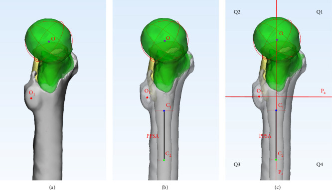
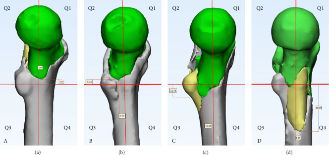
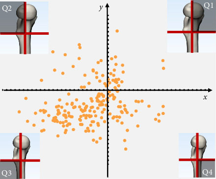
股骨近端内侧壁的完整性对于维持机械稳态至关重要。然而,转子间骨折对内侧壁的影响和最佳诊断方法尚不清楚。我们回顾性分析205例股骨粗隆间骨折患者的CT资料。标记内侧骨折线最低点,建立标准坐标系记录其空间位置。使用Spearman相关分析AO、Evans或Tang分类与不同类型的内侧壁破裂之间的关系。20例患者骨折线最低点位于内侧壁近端第一象限,空间坐标为(6.44±5.47,6.14±2.71)。21例患者位于第二象限,坐标为(-7.23±5.86,8.31±6.59)。122例患者位于第三象限,坐标为(-9.59±4.32,-24.43±15.79);42例患者位于第四象限,坐标为(8.18±4.56,-18.20±12.92)。与AO (r = 0.32, p < 0.001)和Evans (r = 0.38, p < 0.001)分类相比,Tang分类与骨折不稳定性的相关性更强(r = 0.40, p < 0.001)。股骨近端内侧壁在粗隆间骨折中明显受损,其机械稳定性因骨折类型而异。唐氏分类法有效地区分了这些稳定性差异,为临床实践提供了有价值的指导。
Quantitative Analysis of Spatial Distribution of Medial Fracture Lines in Intertrochanteric Fractures.
The integrity of the medial wall of the proximal femur is crucial for maintaining mechanical homeostasis. However, the impact of intertrochanteric fractures on the medial wall and the optimal diagnostic methodology remain unclear. We retrospectively analyzed CT data from 205 patients with intertrochanteric fractures. The lowest point of the medial fracture line was marked, and a standard coordinate system was established to record its spatial position. The association between AO, Evans, or Tang classification and different types of medial wall disruption was analyzed using Spearman correlation. The lowest point of the fracture line was located in the first quadrant of the proximal medial wall in 20 patients, with spatial coordinates of (6.44 ± 5.47, 6.14 ± 2.71). In 21 patients, it was in the second quadrant, with coordinates of (-7.23 ± 5.86, 8.31 ± 6.59). In 122 patients, it was in the third quadrant, with coordinates of (-9.59 ± 4.32, -24.43 ± 15.79), and in 42 patients, it was in the fourth quadrant, with coordinates of (8.18 ± 4.56, -18.20 ± 12.92). The Tang classification showed a stronger correlation with fracture instability (r = 0.40, p < 0.001) compared to the AO (r = 0.32, p < 0.001) and Evans (r = 0.38, p < 0.001) classifications. The medial wall of the proximal femur is significantly compromised in intertrochanteric fractures, with varying mechanical stability depending on the fracture type. The Tang classification effectively differentiates these stability differences, providing valuable guidance for clinical practice.
期刊介绍:
Applied Bionics and Biomechanics publishes papers that seek to understand the mechanics of biological systems, or that use the functions of living organisms as inspiration for the design new devices. Such systems may be used as artificial replacements, or aids, for their original biological purpose, or be used in a different setting altogether.
 求助内容:
求助内容: 应助结果提醒方式:


