基于单光子发射计算机断层扫描/ x射线计算机断层扫描的放射组学分析诊断乳腺癌患者骨转移

IF 3.4 2区 医学 Q2 Medicine
Huiyu Su , Chunwen Ma , Dongli Sun , Jianhua Jin
{"title":"基于单光子发射计算机断层扫描/ x射线计算机断层扫描的放射组学分析诊断乳腺癌患者骨转移","authors":"Huiyu Su ,&nbsp;Chunwen Ma ,&nbsp;Dongli Sun ,&nbsp;Jianhua Jin","doi":"10.1016/j.jbo.2025.100686","DOIUrl":null,"url":null,"abstract":"<div><h3>Purpose</h3><div>Bones are the most metastatic site for breast cancer (BC), which can cause complications such as pathologic osteolysis, seriously affecting the quality of life of patients. This study intended to investigate the efficacy of Single Photon Emission Computed Tomography/X-ray Computed Tomography (SPECT/CT) in diagnosing bone metastases in BC and to develop a model for predicting the diagnostic effectiveness.</div></div><div><h3>Methods</h3><div>In this study, we enrolled 185 patients with BC who underwent SPECT/CT scanning. The region of interest (ROI) of each SPECT/CT image was demarcated, and the radiomics features were determined from the ROIs and screened for the optimal features signature to construct the radiomics model. Based on clinical characteristics, the clinical model was developed, and the independent predictive factors were discovered through univariate and multivariate COX regression analyses. Additionally, the radiomics nomogram was created through integrating the radiomics score and independent predictive factors. Thereafter, the receiver operating characteristic (ROC) was applied to determine the diagnostic performance of various models.</div></div><div><h3>Results</h3><div>The radiomics model was constructed based on 29 optimal features. The N stage was an independent factor, and the radiomics nomogram was created through integrating the radiomics score and N stage. Among three models, the radiomics nomogram had the highest diagnostic value for BC bone metastasis (AUC: the training set: 0.956 (0.909–1.000); the validation set: 0.936 (0.866–1.000)).</div></div><div><h3>Conclusion</h3><div>Radiomics analysis based on SPECT/CT can effectively diagnose bone metastasis in BC patients, establishing a theoretical foundation for the formulation of personalized treatment options in clinical practice.</div></div>","PeriodicalId":48806,"journal":{"name":"Journal of Bone Oncology","volume":"52 ","pages":"Article 100686"},"PeriodicalIF":3.4000,"publicationDate":"2025-05-10","publicationTypes":"Journal Article","fieldsOfStudy":null,"isOpenAccess":false,"openAccessPdf":"","citationCount":"0","resultStr":"{\"title\":\"Single Photon Emission Computed Tomography/X-ray Computed Tomography-based radiomics analysis for diagnosis of bone metastases in patients with breast cancer\",\"authors\":\"Huiyu Su ,&nbsp;Chunwen Ma ,&nbsp;Dongli Sun ,&nbsp;Jianhua Jin\",\"doi\":\"10.1016/j.jbo.2025.100686\",\"DOIUrl\":null,\"url\":null,\"abstract\":\"<div><h3>Purpose</h3><div>Bones are the most metastatic site for breast cancer (BC), which can cause complications such as pathologic osteolysis, seriously affecting the quality of life of patients. This study intended to investigate the efficacy of Single Photon Emission Computed Tomography/X-ray Computed Tomography (SPECT/CT) in diagnosing bone metastases in BC and to develop a model for predicting the diagnostic effectiveness.</div></div><div><h3>Methods</h3><div>In this study, we enrolled 185 patients with BC who underwent SPECT/CT scanning. The region of interest (ROI) of each SPECT/CT image was demarcated, and the radiomics features were determined from the ROIs and screened for the optimal features signature to construct the radiomics model. Based on clinical characteristics, the clinical model was developed, and the independent predictive factors were discovered through univariate and multivariate COX regression analyses. Additionally, the radiomics nomogram was created through integrating the radiomics score and independent predictive factors. Thereafter, the receiver operating characteristic (ROC) was applied to determine the diagnostic performance of various models.</div></div><div><h3>Results</h3><div>The radiomics model was constructed based on 29 optimal features. The N stage was an independent factor, and the radiomics nomogram was created through integrating the radiomics score and N stage. Among three models, the radiomics nomogram had the highest diagnostic value for BC bone metastasis (AUC: the training set: 0.956 (0.909–1.000); the validation set: 0.936 (0.866–1.000)).</div></div><div><h3>Conclusion</h3><div>Radiomics analysis based on SPECT/CT can effectively diagnose bone metastasis in BC patients, establishing a theoretical foundation for the formulation of personalized treatment options in clinical practice.</div></div>\",\"PeriodicalId\":48806,\"journal\":{\"name\":\"Journal of Bone Oncology\",\"volume\":\"52 \",\"pages\":\"Article 100686\"},\"PeriodicalIF\":3.4000,\"publicationDate\":\"2025-05-10\",\"publicationTypes\":\"Journal Article\",\"fieldsOfStudy\":null,\"isOpenAccess\":false,\"openAccessPdf\":\"\",\"citationCount\":\"0\",\"resultStr\":null,\"platform\":\"Semanticscholar\",\"paperid\":null,\"PeriodicalName\":\"Journal of Bone Oncology\",\"FirstCategoryId\":\"3\",\"ListUrlMain\":\"https://www.sciencedirect.com/science/article/pii/S2212137425000272\",\"RegionNum\":2,\"RegionCategory\":\"医学\",\"ArticlePicture\":[],\"TitleCN\":null,\"AbstractTextCN\":null,\"PMCID\":null,\"EPubDate\":\"\",\"PubModel\":\"\",\"JCR\":\"Q2\",\"JCRName\":\"Medicine\",\"Score\":null,\"Total\":0}","platform":"Semanticscholar","paperid":null,"PeriodicalName":"Journal of Bone Oncology","FirstCategoryId":"3","ListUrlMain":"https://www.sciencedirect.com/science/article/pii/S2212137425000272","RegionNum":2,"RegionCategory":"医学","ArticlePicture":[],"TitleCN":null,"AbstractTextCN":null,"PMCID":null,"EPubDate":"","PubModel":"","JCR":"Q2","JCRName":"Medicine","Score":null,"Total":0}
引用次数: 0

摘要

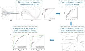
目的:骨是乳腺癌(BC)最易转移的部位,可引起病理性骨溶解等并发症,严重影响患者的生活质量。本研究旨在探讨单光子发射计算机断层扫描/ x射线计算机断层扫描(SPECT/CT)诊断BC骨转移的有效性,并建立预测诊断有效性的模型。方法在本研究中,我们招募了185例接受SPECT/CT扫描的BC患者。对每幅SPECT/CT图像的感兴趣区域(ROI)进行划分,根据感兴趣区域确定放射组学特征,筛选最优特征签名,构建放射组学模型。根据临床特点建立临床模型,通过单因素和多因素COX回归分析发现独立预测因素。此外,通过整合放射组学评分和独立预测因素创建放射组学nomogram。然后,应用受试者工作特征(ROC)来确定各种模型的诊断性能。结果基于29个最优特征构建了放射组学模型。N分期为独立因素,将放射组学评分与N分期相结合,形成放射组学nomogram。三种模型中,放射组学模式图对BC骨转移的诊断价值最高(AUC:训练集:0.956 (0.909 ~ 1.000);验证集:0.936(0.866-1.000))。结论基于SPECT/CT的放射组学分析可有效诊断BC患者骨转移,为临床制定个性化治疗方案奠定理论基础。
本文章由计算机程序翻译,如有差异,请以英文原文为准。
Single Photon Emission Computed Tomography/X-ray Computed Tomography-based radiomics analysis for diagnosis of bone metastases in patients with breast cancer

Purpose

Bones are the most metastatic site for breast cancer (BC), which can cause complications such as pathologic osteolysis, seriously affecting the quality of life of patients. This study intended to investigate the efficacy of Single Photon Emission Computed Tomography/X-ray Computed Tomography (SPECT/CT) in diagnosing bone metastases in BC and to develop a model for predicting the diagnostic effectiveness.

Methods

In this study, we enrolled 185 patients with BC who underwent SPECT/CT scanning. The region of interest (ROI) of each SPECT/CT image was demarcated, and the radiomics features were determined from the ROIs and screened for the optimal features signature to construct the radiomics model. Based on clinical characteristics, the clinical model was developed, and the independent predictive factors were discovered through univariate and multivariate COX regression analyses. Additionally, the radiomics nomogram was created through integrating the radiomics score and independent predictive factors. Thereafter, the receiver operating characteristic (ROC) was applied to determine the diagnostic performance of various models.

Results

The radiomics model was constructed based on 29 optimal features. The N stage was an independent factor, and the radiomics nomogram was created through integrating the radiomics score and N stage. Among three models, the radiomics nomogram had the highest diagnostic value for BC bone metastasis (AUC: the training set: 0.956 (0.909–1.000); the validation set: 0.936 (0.866–1.000)).

Conclusion

Radiomics analysis based on SPECT/CT can effectively diagnose bone metastasis in BC patients, establishing a theoretical foundation for the formulation of personalized treatment options in clinical practice.
求助全文
通过发布文献求助,成功后即可免费获取论文全文。 去求助
来源期刊
CiteScore
7.20
自引率
2.90%
发文量
50
审稿时长
34 days
期刊介绍: The Journal of Bone Oncology is a peer-reviewed international journal aimed at presenting basic, translational and clinical high-quality research related to bone and cancer. As the first journal dedicated to cancer induced bone diseases, JBO welcomes original research articles, review articles, editorials and opinion pieces. Case reports will only be considered in exceptional circumstances and only when accompanied by a comprehensive review of the subject. The areas covered by the journal include: Bone metastases (pathophysiology, epidemiology, diagnostics, clinical features, prevention, treatment) Preclinical models of metastasis Bone microenvironment in cancer (stem cell, bone cell and cancer interactions) Bone targeted therapy (pharmacology, therapeutic targets, drug development, clinical trials, side-effects, outcome research, health economics) Cancer treatment induced bone loss (epidemiology, pathophysiology, prevention and management) Bone imaging (clinical and animal, skeletal interventional radiology) Bone biomarkers (clinical and translational applications) Radiotherapy and radio-isotopes Skeletal complications Bone pain (mechanisms and management) Orthopaedic cancer surgery Primary bone tumours Clinical guidelines Multidisciplinary care Keywords: bisphosphonate, bone, breast cancer, cancer, CTIBL, denosumab, metastasis, myeloma, osteoblast, osteoclast, osteooncology, osteo-oncology, prostate cancer, skeleton, tumour.
×
引用
GB/T 7714-2015
复制
MLA
复制
APA
复制
导出至
BibTeX EndNote RefMan NoteFirst NoteExpress
×
提示
您的信息不完整,为了账户安全,请先补充。
现在去补充
×
提示
您因"违规操作"
具体请查看互助需知
我知道了
×
提示
确定
请完成安全验证×
copy
已复制链接
快去分享给好友吧!
我知道了
右上角分享
点击右上角分享
0
联系我们:info@booksci.cn Book学术提供免费学术资源搜索服务,方便国内外学者检索中英文文献。致力于提供最便捷和优质的服务体验。 Copyright © 2023 布克学术 All rights reserved.
京ICP备2023020795号-1
ghs 京公网安备 11010802042870号
Book学术文献互助
Book学术文献互助群
群 号:481959085
Book学术官方微信