定量数字图像分析评估皮肤纤维化与系统性硬化症患者的组织病理学评分相关

IF 5.1 2区 医学 Q1 MEDICINE, RESEARCH & EXPERIMENTAL
Ruben Oganesyan , Dimitra Pouli , Bo Shi , Mary Carns , Kathleen Dennis-Aren , Maria E. Teves , John Varga , Rosalynn M. Nazarian
{"title":"定量数字图像分析评估皮肤纤维化与系统性硬化症患者的组织病理学评分相关","authors":"Ruben Oganesyan ,&nbsp;Dimitra Pouli ,&nbsp;Bo Shi ,&nbsp;Mary Carns ,&nbsp;Kathleen Dennis-Aren ,&nbsp;Maria E. Teves ,&nbsp;John Varga ,&nbsp;Rosalynn M. Nazarian","doi":"10.1016/j.labinv.2025.104177","DOIUrl":null,"url":null,"abstract":"<div><div>Systemic sclerosis (SSc) is a multisystem autoimmune disease characterized by progressive fibrosis of the skin, blood vessels, and internal organs. Accurate assessment of skin fibrosis is essential for disease monitoring and treatment evaluation, yet reliable quantitative methods are lacking. We compared computer-assisted digital image analysis with traditional semiquantitative histopathological scoring. Patients with SSc and healthy controls were identified from a hospital database. Clinical data, modified Rodnan skin score, and hematoxylin and eosin–stained skin biopsies were collected. Biopsies were scored using modified Keyser and Farge criteria. Whole-slide images were analyzed using QuPath open source digital pathology software (v0.5.1) for area annotation, normalized cellularity, and collagen density, whereas collagen alignment was evaluated using CurveAlign (MATLAB) software. Seven SSc and 7 healthy controls (no significant demographic differences) were studied. SSc samples showed significant differences in hyalinized collagen (<em>P</em> &lt; .001), dermal fibroblast cellularity (<em>P</em> = .009), and total histopathological score (<em>P</em> = .007). Quantitative analysis confirmed decreased dermal cellularity (<em>P</em> = .024), increased collagen density (<em>P</em> = .009), and higher collagen alignment, with reduced fiber orientation variability, reflecting extracellular matrix restructuring in SSc. Tools such as QuPath and CurveAlign improve objectivity and reproducibility in SSc skin assessment, correlating well with traditional scores. These findings support the integration of computer-aided quantitative analysis into clinical practice and trials, advancing personalized fibrosis evaluation. Larger, longitudinal studies are needed for further validation.</div></div>","PeriodicalId":17930,"journal":{"name":"Laboratory Investigation","volume":"105 8","pages":"Article 104177"},"PeriodicalIF":5.1000,"publicationDate":"2025-04-20","publicationTypes":"Journal Article","fieldsOfStudy":null,"isOpenAccess":false,"openAccessPdf":"","citationCount":"0","resultStr":"{\"title\":\"Quantitative Digital Image Analysis for Assessing Cutaneous Fibrosis Correlates With Histopathological Scoring in Patients With Systemic Sclerosis\",\"authors\":\"Ruben Oganesyan ,&nbsp;Dimitra Pouli ,&nbsp;Bo Shi ,&nbsp;Mary Carns ,&nbsp;Kathleen Dennis-Aren ,&nbsp;Maria E. Teves ,&nbsp;John Varga ,&nbsp;Rosalynn M. Nazarian\",\"doi\":\"10.1016/j.labinv.2025.104177\",\"DOIUrl\":null,\"url\":null,\"abstract\":\"<div><div>Systemic sclerosis (SSc) is a multisystem autoimmune disease characterized by progressive fibrosis of the skin, blood vessels, and internal organs. Accurate assessment of skin fibrosis is essential for disease monitoring and treatment evaluation, yet reliable quantitative methods are lacking. We compared computer-assisted digital image analysis with traditional semiquantitative histopathological scoring. Patients with SSc and healthy controls were identified from a hospital database. Clinical data, modified Rodnan skin score, and hematoxylin and eosin–stained skin biopsies were collected. Biopsies were scored using modified Keyser and Farge criteria. Whole-slide images were analyzed using QuPath open source digital pathology software (v0.5.1) for area annotation, normalized cellularity, and collagen density, whereas collagen alignment was evaluated using CurveAlign (MATLAB) software. Seven SSc and 7 healthy controls (no significant demographic differences) were studied. SSc samples showed significant differences in hyalinized collagen (<em>P</em> &lt; .001), dermal fibroblast cellularity (<em>P</em> = .009), and total histopathological score (<em>P</em> = .007). Quantitative analysis confirmed decreased dermal cellularity (<em>P</em> = .024), increased collagen density (<em>P</em> = .009), and higher collagen alignment, with reduced fiber orientation variability, reflecting extracellular matrix restructuring in SSc. Tools such as QuPath and CurveAlign improve objectivity and reproducibility in SSc skin assessment, correlating well with traditional scores. These findings support the integration of computer-aided quantitative analysis into clinical practice and trials, advancing personalized fibrosis evaluation. Larger, longitudinal studies are needed for further validation.</div></div>\",\"PeriodicalId\":17930,\"journal\":{\"name\":\"Laboratory Investigation\",\"volume\":\"105 8\",\"pages\":\"Article 104177\"},\"PeriodicalIF\":5.1000,\"publicationDate\":\"2025-04-20\",\"publicationTypes\":\"Journal Article\",\"fieldsOfStudy\":null,\"isOpenAccess\":false,\"openAccessPdf\":\"\",\"citationCount\":\"0\",\"resultStr\":null,\"platform\":\"Semanticscholar\",\"paperid\":null,\"PeriodicalName\":\"Laboratory Investigation\",\"FirstCategoryId\":\"3\",\"ListUrlMain\":\"https://www.sciencedirect.com/science/article/pii/S002368372500087X\",\"RegionNum\":2,\"RegionCategory\":\"医学\",\"ArticlePicture\":[],\"TitleCN\":null,\"AbstractTextCN\":null,\"PMCID\":null,\"EPubDate\":\"\",\"PubModel\":\"\",\"JCR\":\"Q1\",\"JCRName\":\"MEDICINE, RESEARCH & EXPERIMENTAL\",\"Score\":null,\"Total\":0}","platform":"Semanticscholar","paperid":null,"PeriodicalName":"Laboratory Investigation","FirstCategoryId":"3","ListUrlMain":"https://www.sciencedirect.com/science/article/pii/S002368372500087X","RegionNum":2,"RegionCategory":"医学","ArticlePicture":[],"TitleCN":null,"AbstractTextCN":null,"PMCID":null,"EPubDate":"","PubModel":"","JCR":"Q1","JCRName":"MEDICINE, RESEARCH & EXPERIMENTAL","Score":null,"Total":0}
引用次数: 0

摘要

系统性硬化症(SSc)是一种以皮肤、血管和内脏器官进行性纤维化为特征的多系统自身免疫性疾病。准确评估皮肤纤维化对疾病监测和治疗评估至关重要,但缺乏可靠的定量方法。我们比较了计算机辅助的数字图像分析与传统的半定量组织病理学评分。从医院数据库中确定SSc患者和健康对照者。收集临床资料、改良罗德曼皮肤评分、苏木精和伊红染色皮肤活检。活检采用改良的Keyser和Farge标准评分。使用QuPath开源数字病理软件(v0.5.1)分析全片图像,进行区域注释、归一化细胞度和胶原蛋白密度分析,而使用CurveAlign (MATLAB)软件评估胶原蛋白排列。研究了7名SSc和7名健康对照(无显著人口统计学差异)。SSc样品在透明化胶原蛋白(P <;.001)、真皮成纤维细胞结构(P = .009)和总组织病理学评分(P = .007)。定量分析证实真皮细胞数量减少(P = 0.024),胶原密度增加(P = 0.009),胶原排列更高,纤维取向变异性减少,反映了SSc的细胞外基质重构。QuPath和CurveAlign等工具提高了SSc皮肤评估的客观性和可重复性,与传统评分有很好的相关性。这些发现支持将计算机辅助定量分析整合到临床实践和试验中,推进个性化纤维化评估。需要更大规模的纵向研究来进一步验证。
本文章由计算机程序翻译,如有差异,请以英文原文为准。
Quantitative Digital Image Analysis for Assessing Cutaneous Fibrosis Correlates With Histopathological Scoring in Patients With Systemic Sclerosis
Systemic sclerosis (SSc) is a multisystem autoimmune disease characterized by progressive fibrosis of the skin, blood vessels, and internal organs. Accurate assessment of skin fibrosis is essential for disease monitoring and treatment evaluation, yet reliable quantitative methods are lacking. We compared computer-assisted digital image analysis with traditional semiquantitative histopathological scoring. Patients with SSc and healthy controls were identified from a hospital database. Clinical data, modified Rodnan skin score, and hematoxylin and eosin–stained skin biopsies were collected. Biopsies were scored using modified Keyser and Farge criteria. Whole-slide images were analyzed using QuPath open source digital pathology software (v0.5.1) for area annotation, normalized cellularity, and collagen density, whereas collagen alignment was evaluated using CurveAlign (MATLAB) software. Seven SSc and 7 healthy controls (no significant demographic differences) were studied. SSc samples showed significant differences in hyalinized collagen (P < .001), dermal fibroblast cellularity (P = .009), and total histopathological score (P = .007). Quantitative analysis confirmed decreased dermal cellularity (P = .024), increased collagen density (P = .009), and higher collagen alignment, with reduced fiber orientation variability, reflecting extracellular matrix restructuring in SSc. Tools such as QuPath and CurveAlign improve objectivity and reproducibility in SSc skin assessment, correlating well with traditional scores. These findings support the integration of computer-aided quantitative analysis into clinical practice and trials, advancing personalized fibrosis evaluation. Larger, longitudinal studies are needed for further validation.
求助全文
通过发布文献求助,成功后即可免费获取论文全文。 去求助
来源期刊
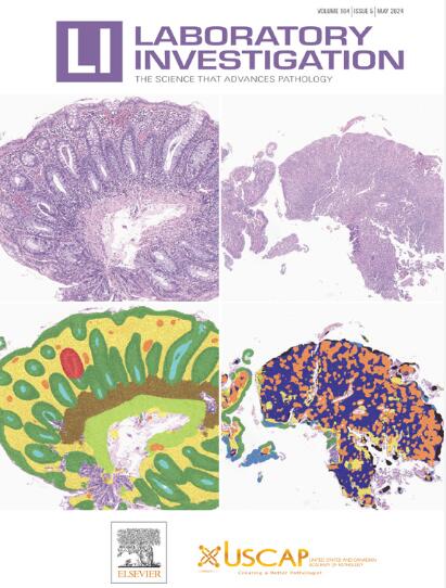
Laboratory Investigation
Laboratory Investigation 医学-病理学
CiteScore
8.30
自引率
0.00%
发文量
125
审稿时长
2 months
期刊介绍: Laboratory Investigation is an international journal owned by the United States and Canadian Academy of Pathology. Laboratory Investigation offers prompt publication of high-quality original research in all biomedical disciplines relating to the understanding of human disease and the application of new methods to the diagnosis of disease. Both human and experimental studies are welcome.
×
引用
GB/T 7714-2015
复制
MLA
复制
APA
复制
导出至
BibTeX EndNote RefMan NoteFirst NoteExpress
×
提示
您的信息不完整,为了账户安全,请先补充。
现在去补充
×
提示
您因"违规操作"
具体请查看互助需知
我知道了
×
提示
确定
请完成安全验证×
copy
已复制链接
快去分享给好友吧!
我知道了
右上角分享
点击右上角分享
0
联系我们:info@booksci.cn Book学术提供免费学术资源搜索服务,方便国内外学者检索中英文文献。致力于提供最便捷和优质的服务体验。 Copyright © 2023 布克学术 All rights reserved.
京ICP备2023020795号-1
ghs 京公网安备 11010802042870号
Book学术文献互助
Book学术文献互助群
群 号:481959085
Book学术官方微信