Matthew Sinovich, Josep Monné Rodriguez, Aldona Pieńkowska-Schelling, Claude Schelling, Padraig G Kelly
{"title":"罕见的蒙兰马腹部保留睾丸的病例。","authors":"Matthew Sinovich, Josep Monné Rodriguez, Aldona Pieńkowska-Schelling, Claude Schelling, Padraig G Kelly","doi":"10.1159/000545559","DOIUrl":null,"url":null,"abstract":"<p><strong>Introduction: </strong>Monorchidism is a rarely described condition in the horse and is not to be confused with cryptorchidism. The diagnosis is challenging and confirmed by surgery and histology in combination with hormonal assays. This report describes, to the best of the author's knowledge, the first case of monorchidism and abdominal cryptorchidism of the developed testicle in a horse.</p><p><strong>Methods: </strong>An Irish Cob underwent laparoscopic castration for removal of bilateral cryptorchid testicles. At surgery, the horse was diagnosed as a monorchid with the testicle retained intra-abdominally. Histopathological, hormonal, molecular and cytogenetic analysis was performed. This included measuring testosterone and anti-Mullerian hormone (AMH) in serum blood, isolating genomic DNA from EDTA- and heparin-treated blood, PCR amplification of the SRY gene, metaphase chromosome preparation, and DAPI banding before metaphase analysis with fluorescence in situ hybridisation (FISH) analysis.</p><p><strong>Results: </strong>The horse was positive for the SRY gene and had a mosaic 63,X/64,XY karyotype with the aneuploid cells being present in only 2% of metaphases. FISH showed that the missing sex chromosome of the aneuploid cell line was the Y chromosome embedded in micronuclei. An abnormal high rate of micronuclei (6.6%) was observed indicating genotoxic events and/or genome instability. Hormonal assay results confirmed that AMH was not significantly increased, suggesting that no further testicular tissue was present. Histopathology was consistent with testicular tissue displaying a Sertoli cell-only pattern with bipolar ductal structures.</p><p><strong>Conclusion: </strong>The exact causes of monorchidism and cryptorchidism are unclear, but the elevated rate of micronuclei is clear evidence for genome instability which might have been involved in the failure of normal testicular development and descent. Future cases could further clarify the disease mechanism based on this report.</p>","PeriodicalId":49536,"journal":{"name":"Sexual Development","volume":" ","pages":"1-9"},"PeriodicalIF":2.4000,"publicationDate":"2025-04-08","publicationTypes":"Journal Article","fieldsOfStudy":null,"isOpenAccess":false,"openAccessPdf":"https://www.ncbi.nlm.nih.gov/pmc/articles/PMC12119058/pdf/","citationCount":"0","resultStr":"{\"title\":\"An Unusual Case of a Monorchid Horse with an Abdominally Retained Testicle.\",\"authors\":\"Matthew Sinovich, Josep Monné Rodriguez, Aldona Pieńkowska-Schelling, Claude Schelling, Padraig G Kelly\",\"doi\":\"10.1159/000545559\",\"DOIUrl\":null,\"url\":null,\"abstract\":\"<p><strong>Introduction: </strong>Monorchidism is a rarely described condition in the horse and is not to be confused with cryptorchidism. The diagnosis is challenging and confirmed by surgery and histology in combination with hormonal assays. This report describes, to the best of the author's knowledge, the first case of monorchidism and abdominal cryptorchidism of the developed testicle in a horse.</p><p><strong>Methods: </strong>An Irish Cob underwent laparoscopic castration for removal of bilateral cryptorchid testicles. At surgery, the horse was diagnosed as a monorchid with the testicle retained intra-abdominally. Histopathological, hormonal, molecular and cytogenetic analysis was performed. This included measuring testosterone and anti-Mullerian hormone (AMH) in serum blood, isolating genomic DNA from EDTA- and heparin-treated blood, PCR amplification of the SRY gene, metaphase chromosome preparation, and DAPI banding before metaphase analysis with fluorescence in situ hybridisation (FISH) analysis.</p><p><strong>Results: </strong>The horse was positive for the SRY gene and had a mosaic 63,X/64,XY karyotype with the aneuploid cells being present in only 2% of metaphases. FISH showed that the missing sex chromosome of the aneuploid cell line was the Y chromosome embedded in micronuclei. An abnormal high rate of micronuclei (6.6%) was observed indicating genotoxic events and/or genome instability. Hormonal assay results confirmed that AMH was not significantly increased, suggesting that no further testicular tissue was present. Histopathology was consistent with testicular tissue displaying a Sertoli cell-only pattern with bipolar ductal structures.</p><p><strong>Conclusion: </strong>The exact causes of monorchidism and cryptorchidism are unclear, but the elevated rate of micronuclei is clear evidence for genome instability which might have been involved in the failure of normal testicular development and descent. Future cases could further clarify the disease mechanism based on this report.</p>\",\"PeriodicalId\":49536,\"journal\":{\"name\":\"Sexual Development\",\"volume\":\" \",\"pages\":\"1-9\"},\"PeriodicalIF\":2.4000,\"publicationDate\":\"2025-04-08\",\"publicationTypes\":\"Journal Article\",\"fieldsOfStudy\":null,\"isOpenAccess\":false,\"openAccessPdf\":\"https://www.ncbi.nlm.nih.gov/pmc/articles/PMC12119058/pdf/\",\"citationCount\":\"0\",\"resultStr\":null,\"platform\":\"Semanticscholar\",\"paperid\":null,\"PeriodicalName\":\"Sexual Development\",\"FirstCategoryId\":\"3\",\"ListUrlMain\":\"https://doi.org/10.1159/000545559\",\"RegionNum\":4,\"RegionCategory\":\"医学\",\"ArticlePicture\":[],\"TitleCN\":null,\"AbstractTextCN\":null,\"PMCID\":null,\"EPubDate\":\"\",\"PubModel\":\"\",\"JCR\":\"Q2\",\"JCRName\":\"DEVELOPMENTAL BIOLOGY\",\"Score\":null,\"Total\":0}","platform":"Semanticscholar","paperid":null,"PeriodicalName":"Sexual Development","FirstCategoryId":"3","ListUrlMain":"https://doi.org/10.1159/000545559","RegionNum":4,"RegionCategory":"医学","ArticlePicture":[],"TitleCN":null,"AbstractTextCN":null,"PMCID":null,"EPubDate":"","PubModel":"","JCR":"Q2","JCRName":"DEVELOPMENTAL BIOLOGY","Score":null,"Total":0}
An Unusual Case of a Monorchid Horse with an Abdominally Retained Testicle.
Introduction: Monorchidism is a rarely described condition in the horse and is not to be confused with cryptorchidism. The diagnosis is challenging and confirmed by surgery and histology in combination with hormonal assays. This report describes, to the best of the author's knowledge, the first case of monorchidism and abdominal cryptorchidism of the developed testicle in a horse.
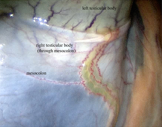
Methods: An Irish Cob underwent laparoscopic castration for removal of bilateral cryptorchid testicles. At surgery, the horse was diagnosed as a monorchid with the testicle retained intra-abdominally. Histopathological, hormonal, molecular and cytogenetic analysis was performed. This included measuring testosterone and anti-Mullerian hormone (AMH) in serum blood, isolating genomic DNA from EDTA- and heparin-treated blood, PCR amplification of the SRY gene, metaphase chromosome preparation, and DAPI banding before metaphase analysis with fluorescence in situ hybridisation (FISH) analysis.
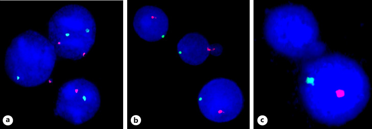
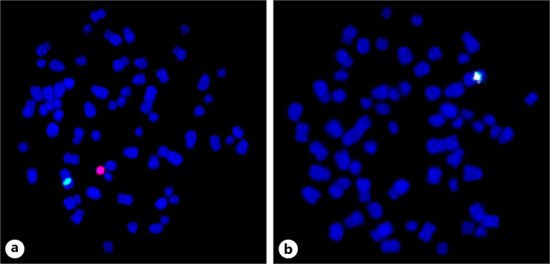
Results: The horse was positive for the SRY gene and had a mosaic 63,X/64,XY karyotype with the aneuploid cells being present in only 2% of metaphases. FISH showed that the missing sex chromosome of the aneuploid cell line was the Y chromosome embedded in micronuclei. An abnormal high rate of micronuclei (6.6%) was observed indicating genotoxic events and/or genome instability. Hormonal assay results confirmed that AMH was not significantly increased, suggesting that no further testicular tissue was present. Histopathology was consistent with testicular tissue displaying a Sertoli cell-only pattern with bipolar ductal structures.
Conclusion: The exact causes of monorchidism and cryptorchidism are unclear, but the elevated rate of micronuclei is clear evidence for genome instability which might have been involved in the failure of normal testicular development and descent. Future cases could further clarify the disease mechanism based on this report.
期刊介绍:
Recent discoveries in experimental and clinical research have led to impressive advances in our knowledge of the genetic and environmental mechanisms governing sex determination and differentiation, their evolution as well as the mutations or endocrine and metabolic abnormalities that interfere with normal gonadal development. ‘Sexual Development’ provides a unique forum for this rapidly expanding field. Its broad scope covers all aspects of genetics, molecular biology, embryology, endocrinology, evolution and pathology of sex determination and differentiation in humans and animals. It publishes high-quality original research manuscripts, review articles, short reports, case reports and commentaries. An internationally renowned and multidisciplinary editorial team of three chief editors, ten prominent scientists serving as section editors, and a distinguished panel of editorial board members ensures fast and author-friendly editorial processing and peer reviewing.
 求助内容:
求助内容: 应助结果提醒方式:


