{"title":"一只实验用小猎犬患有伴有肿瘤细胞改变的淋巴细胞性甲状腺炎。","authors":"Osamu Hashiguchi, Kohji Tanaka, Yuko Yamaguchi, Moeko Aoki, Nobuaki Sato, Takuro Endo, Maoko Yamaguchi, Tsubasa Saito","doi":"10.1293/tox.2024-0073","DOIUrl":null,"url":null,"abstract":"<p><p>Histopathological, immunohistochemical, and ultrastructural characteristics of lymphocytic thyroiditis in an untreated four-year-old male beagle were described. Histopathologically, the thyroid glands were composed of two distinct cell types: round to oval cells with eosinophilic granular cytoplasm (Type A), which is consistent with the features of oncocytic oxyphils, and larger round cells with amphophilic or pale cytoplasm (Type B). These cell types extensively and diffusely infiltrated with a mixture of lymphocytes and plasma cells, destroying the follicular structure. Immunohistochemistry revealed that Type A cells were positive for thyroglobulin and cytochrome C, and that Type B cells were positive for calcitonin, synaptophysin, and cytochrome C. These results indicate that Type A and B cells stem from follicular and C cells, respectively. Ultrastructural investigation showed that microfollicles and microvilli were evident in the cytoplasm and along the luminal surface of Type A cells. Thus, the lymphocytic thyroiditis observed in the beagle exhibited a morphology similar to that of Hashimoto thyroiditis in humans, particularly in view of an oncocytic alteration of follicular cells.</p>","PeriodicalId":17437,"journal":{"name":"Journal of Toxicologic Pathology","volume":"38 2","pages":"177-182"},"PeriodicalIF":0.9000,"publicationDate":"2025-04-01","publicationTypes":"Journal Article","fieldsOfStudy":null,"isOpenAccess":false,"openAccessPdf":"https://www.ncbi.nlm.nih.gov/pmc/articles/PMC11966123/pdf/","citationCount":"0","resultStr":"{\"title\":\"Lymphocytic thyroiditis with an oncocytic alteration in a laboratory beagle.\",\"authors\":\"Osamu Hashiguchi, Kohji Tanaka, Yuko Yamaguchi, Moeko Aoki, Nobuaki Sato, Takuro Endo, Maoko Yamaguchi, Tsubasa Saito\",\"doi\":\"10.1293/tox.2024-0073\",\"DOIUrl\":null,\"url\":null,\"abstract\":\"<p><p>Histopathological, immunohistochemical, and ultrastructural characteristics of lymphocytic thyroiditis in an untreated four-year-old male beagle were described. Histopathologically, the thyroid glands were composed of two distinct cell types: round to oval cells with eosinophilic granular cytoplasm (Type A), which is consistent with the features of oncocytic oxyphils, and larger round cells with amphophilic or pale cytoplasm (Type B). These cell types extensively and diffusely infiltrated with a mixture of lymphocytes and plasma cells, destroying the follicular structure. Immunohistochemistry revealed that Type A cells were positive for thyroglobulin and cytochrome C, and that Type B cells were positive for calcitonin, synaptophysin, and cytochrome C. These results indicate that Type A and B cells stem from follicular and C cells, respectively. Ultrastructural investigation showed that microfollicles and microvilli were evident in the cytoplasm and along the luminal surface of Type A cells. Thus, the lymphocytic thyroiditis observed in the beagle exhibited a morphology similar to that of Hashimoto thyroiditis in humans, particularly in view of an oncocytic alteration of follicular cells.</p>\",\"PeriodicalId\":17437,\"journal\":{\"name\":\"Journal of Toxicologic Pathology\",\"volume\":\"38 2\",\"pages\":\"177-182\"},\"PeriodicalIF\":0.9000,\"publicationDate\":\"2025-04-01\",\"publicationTypes\":\"Journal Article\",\"fieldsOfStudy\":null,\"isOpenAccess\":false,\"openAccessPdf\":\"https://www.ncbi.nlm.nih.gov/pmc/articles/PMC11966123/pdf/\",\"citationCount\":\"0\",\"resultStr\":null,\"platform\":\"Semanticscholar\",\"paperid\":null,\"PeriodicalName\":\"Journal of Toxicologic Pathology\",\"FirstCategoryId\":\"3\",\"ListUrlMain\":\"https://doi.org/10.1293/tox.2024-0073\",\"RegionNum\":4,\"RegionCategory\":\"医学\",\"ArticlePicture\":[],\"TitleCN\":null,\"AbstractTextCN\":null,\"PMCID\":null,\"EPubDate\":\"2024/12/30 0:00:00\",\"PubModel\":\"Epub\",\"JCR\":\"Q4\",\"JCRName\":\"PATHOLOGY\",\"Score\":null,\"Total\":0}","platform":"Semanticscholar","paperid":null,"PeriodicalName":"Journal of Toxicologic Pathology","FirstCategoryId":"3","ListUrlMain":"https://doi.org/10.1293/tox.2024-0073","RegionNum":4,"RegionCategory":"医学","ArticlePicture":[],"TitleCN":null,"AbstractTextCN":null,"PMCID":null,"EPubDate":"2024/12/30 0:00:00","PubModel":"Epub","JCR":"Q4","JCRName":"PATHOLOGY","Score":null,"Total":0}
Lymphocytic thyroiditis with an oncocytic alteration in a laboratory beagle.
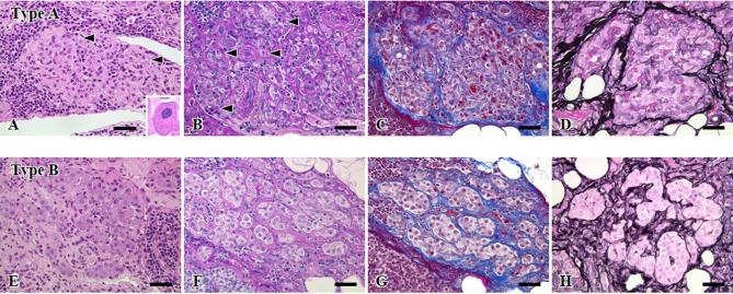
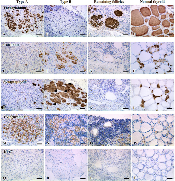
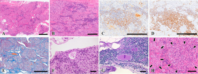
Histopathological, immunohistochemical, and ultrastructural characteristics of lymphocytic thyroiditis in an untreated four-year-old male beagle were described. Histopathologically, the thyroid glands were composed of two distinct cell types: round to oval cells with eosinophilic granular cytoplasm (Type A), which is consistent with the features of oncocytic oxyphils, and larger round cells with amphophilic or pale cytoplasm (Type B). These cell types extensively and diffusely infiltrated with a mixture of lymphocytes and plasma cells, destroying the follicular structure. Immunohistochemistry revealed that Type A cells were positive for thyroglobulin and cytochrome C, and that Type B cells were positive for calcitonin, synaptophysin, and cytochrome C. These results indicate that Type A and B cells stem from follicular and C cells, respectively. Ultrastructural investigation showed that microfollicles and microvilli were evident in the cytoplasm and along the luminal surface of Type A cells. Thus, the lymphocytic thyroiditis observed in the beagle exhibited a morphology similar to that of Hashimoto thyroiditis in humans, particularly in view of an oncocytic alteration of follicular cells.
期刊介绍:
JTP is a scientific journal that publishes original studies in the field of toxicological pathology and in a wide variety of other related fields. The main scope of the journal is listed below.
Administrative Opinions of Policymakers and Regulatory Agencies
Adverse Events
Carcinogenesis
Data of A Predominantly Negative Nature
Drug-Induced Hematologic Toxicity
Embryological Pathology
High Throughput Pathology
Historical Data of Experimental Animals
Immunohistochemical Analysis
Molecular Pathology
Nomenclature of Lesions
Non-mammal Toxicity Study
Result or Lesion Induced by Chemicals of Which Names Hidden on Account of the Authors
Technology and Methodology Related to Toxicological Pathology
Tumor Pathology; Neoplasia and Hyperplasia
Ultrastructural Analysis
Use of Animal Models.
 求助内容:
求助内容: 应助结果提醒方式:


