Nico Sollmann, Kai Mei, Maximilian T Löffler, Sebastian Rühling, Meinrad Beer, Claus Zimmer, Jan S Kirschke, Peter B Noël, Thomas Baum, Julio Carballido-Gamio
{"title":"模拟低剂量多探测器计算机断层扫描:股骨近端骨强度替代参数的空间效应。","authors":"Nico Sollmann, Kai Mei, Maximilian T Löffler, Sebastian Rühling, Meinrad Beer, Claus Zimmer, Jan S Kirschke, Peter B Noël, Thomas Baum, Julio Carballido-Gamio","doi":"10.1007/s00198-025-07467-4","DOIUrl":null,"url":null,"abstract":"<p><p>This study investigated simulated tube current reduction and sparse sampling for low-dose computed tomography (CT) regarding volumetric bone mineral density (vBMD) and cortical bone thickness (Ct.Th) of the proximal femur. Sparse sampling with dose reductions of up to 90% may still allow extraction of bone strength parameters with clinically acceptable accuracy.</p><p><strong>Introduction: </strong>We aimed to investigate effects of CT with simulated lowered tube current and sparse sampling on trabecular and cortical vBMD as well as Ct.Th of the entire proximal femur, its subregions, and with detailed spatial assessments.</p><p><strong>Methods: </strong>Clinical routine multi-detector CT (MDCT) scans covering the hips from 40 patients were used for simulations of low-dose imaging with 50% and 10% of the original tube current (D50, D10) or projections (P50, P10) combined with statistical iterative reconstruction (SIR), which were then compared against original data with full dose (D100 P100) regarding trabecular vBMD, cortical vBMD, and Ct.Th. An automated framework for multi-parametric assessments was used. Relative errors by comparing measures from original data and simulated low-dose data, regression analyses, Bland-Altman analyses, and statistical parametric mapping (SPM, to assess the spatial distribution of accuracy) were computed.</p><p><strong>Results: </strong>Sparse sampling enabled drastic reductions of radiation exposure (down to 10% of original imaging) while still producing determinants of bone strength with clinically acceptable relative changes. Lower biases according to Bland-Altman analyses were observed for sparse sampling compared to imaging with virtually lowered tube currents (D10 P100 versus D100 P10) regarding trabecular vBMD, cortical vBMD, as well as Ct.Th. Better accuracy across the whole proximal femur for D100 P50 than for D50 P100 and for D100 P10 than for D10 P100 was observed.</p><p><strong>Conclusions: </strong>Sparse sampling with SIR may enable drastic reductions of radiation exposure (up to 90% of original doses) for opportunistically measuring image-based surrogate parameters of bone strength.</p>","PeriodicalId":19638,"journal":{"name":"Osteoporosis International","volume":" ","pages":"917-928"},"PeriodicalIF":5.4000,"publicationDate":"2025-05-01","publicationTypes":"Journal Article","fieldsOfStudy":null,"isOpenAccess":false,"openAccessPdf":"https://www.ncbi.nlm.nih.gov/pmc/articles/PMC12089198/pdf/","citationCount":"0","resultStr":"{\"title\":\"Simulated low-dose multi-detector computed tomography: spatial effects on surrogate parameters of bone strength at the proximal femur.\",\"authors\":\"Nico Sollmann, Kai Mei, Maximilian T Löffler, Sebastian Rühling, Meinrad Beer, Claus Zimmer, Jan S Kirschke, Peter B Noël, Thomas Baum, Julio Carballido-Gamio\",\"doi\":\"10.1007/s00198-025-07467-4\",\"DOIUrl\":null,\"url\":null,\"abstract\":\"<p><p>This study investigated simulated tube current reduction and sparse sampling for low-dose computed tomography (CT) regarding volumetric bone mineral density (vBMD) and cortical bone thickness (Ct.Th) of the proximal femur. Sparse sampling with dose reductions of up to 90% may still allow extraction of bone strength parameters with clinically acceptable accuracy.</p><p><strong>Introduction: </strong>We aimed to investigate effects of CT with simulated lowered tube current and sparse sampling on trabecular and cortical vBMD as well as Ct.Th of the entire proximal femur, its subregions, and with detailed spatial assessments.</p><p><strong>Methods: </strong>Clinical routine multi-detector CT (MDCT) scans covering the hips from 40 patients were used for simulations of low-dose imaging with 50% and 10% of the original tube current (D50, D10) or projections (P50, P10) combined with statistical iterative reconstruction (SIR), which were then compared against original data with full dose (D100 P100) regarding trabecular vBMD, cortical vBMD, and Ct.Th. An automated framework for multi-parametric assessments was used. Relative errors by comparing measures from original data and simulated low-dose data, regression analyses, Bland-Altman analyses, and statistical parametric mapping (SPM, to assess the spatial distribution of accuracy) were computed.</p><p><strong>Results: </strong>Sparse sampling enabled drastic reductions of radiation exposure (down to 10% of original imaging) while still producing determinants of bone strength with clinically acceptable relative changes. Lower biases according to Bland-Altman analyses were observed for sparse sampling compared to imaging with virtually lowered tube currents (D10 P100 versus D100 P10) regarding trabecular vBMD, cortical vBMD, as well as Ct.Th. Better accuracy across the whole proximal femur for D100 P50 than for D50 P100 and for D100 P10 than for D10 P100 was observed.</p><p><strong>Conclusions: </strong>Sparse sampling with SIR may enable drastic reductions of radiation exposure (up to 90% of original doses) for opportunistically measuring image-based surrogate parameters of bone strength.</p>\",\"PeriodicalId\":19638,\"journal\":{\"name\":\"Osteoporosis International\",\"volume\":\" \",\"pages\":\"917-928\"},\"PeriodicalIF\":5.4000,\"publicationDate\":\"2025-05-01\",\"publicationTypes\":\"Journal Article\",\"fieldsOfStudy\":null,\"isOpenAccess\":false,\"openAccessPdf\":\"https://www.ncbi.nlm.nih.gov/pmc/articles/PMC12089198/pdf/\",\"citationCount\":\"0\",\"resultStr\":null,\"platform\":\"Semanticscholar\",\"paperid\":null,\"PeriodicalName\":\"Osteoporosis International\",\"FirstCategoryId\":\"3\",\"ListUrlMain\":\"https://doi.org/10.1007/s00198-025-07467-4\",\"RegionNum\":2,\"RegionCategory\":\"医学\",\"ArticlePicture\":[],\"TitleCN\":null,\"AbstractTextCN\":null,\"PMCID\":null,\"EPubDate\":\"2025/4/5 0:00:00\",\"PubModel\":\"Epub\",\"JCR\":\"Q1\",\"JCRName\":\"ENDOCRINOLOGY & METABOLISM\",\"Score\":null,\"Total\":0}","platform":"Semanticscholar","paperid":null,"PeriodicalName":"Osteoporosis International","FirstCategoryId":"3","ListUrlMain":"https://doi.org/10.1007/s00198-025-07467-4","RegionNum":2,"RegionCategory":"医学","ArticlePicture":[],"TitleCN":null,"AbstractTextCN":null,"PMCID":null,"EPubDate":"2025/4/5 0:00:00","PubModel":"Epub","JCR":"Q1","JCRName":"ENDOCRINOLOGY & METABOLISM","Score":null,"Total":0}
Simulated low-dose multi-detector computed tomography: spatial effects on surrogate parameters of bone strength at the proximal femur.
This study investigated simulated tube current reduction and sparse sampling for low-dose computed tomography (CT) regarding volumetric bone mineral density (vBMD) and cortical bone thickness (Ct.Th) of the proximal femur. Sparse sampling with dose reductions of up to 90% may still allow extraction of bone strength parameters with clinically acceptable accuracy.
Introduction: We aimed to investigate effects of CT with simulated lowered tube current and sparse sampling on trabecular and cortical vBMD as well as Ct.Th of the entire proximal femur, its subregions, and with detailed spatial assessments.
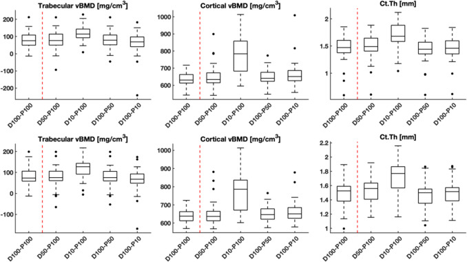
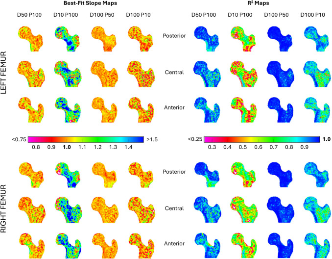
Methods: Clinical routine multi-detector CT (MDCT) scans covering the hips from 40 patients were used for simulations of low-dose imaging with 50% and 10% of the original tube current (D50, D10) or projections (P50, P10) combined with statistical iterative reconstruction (SIR), which were then compared against original data with full dose (D100 P100) regarding trabecular vBMD, cortical vBMD, and Ct.Th. An automated framework for multi-parametric assessments was used. Relative errors by comparing measures from original data and simulated low-dose data, regression analyses, Bland-Altman analyses, and statistical parametric mapping (SPM, to assess the spatial distribution of accuracy) were computed.
Results: Sparse sampling enabled drastic reductions of radiation exposure (down to 10% of original imaging) while still producing determinants of bone strength with clinically acceptable relative changes. Lower biases according to Bland-Altman analyses were observed for sparse sampling compared to imaging with virtually lowered tube currents (D10 P100 versus D100 P10) regarding trabecular vBMD, cortical vBMD, as well as Ct.Th. Better accuracy across the whole proximal femur for D100 P50 than for D50 P100 and for D100 P10 than for D10 P100 was observed.
Conclusions: Sparse sampling with SIR may enable drastic reductions of radiation exposure (up to 90% of original doses) for opportunistically measuring image-based surrogate parameters of bone strength.
期刊介绍:
An international multi-disciplinary journal which is a joint initiative between the International Osteoporosis Foundation and the National Osteoporosis Foundation of the USA, Osteoporosis International provides a forum for the communication and exchange of current ideas concerning the diagnosis, prevention, treatment and management of osteoporosis and other metabolic bone diseases.
It publishes: original papers - reporting progress and results in all areas of osteoporosis and its related fields; review articles - reflecting the present state of knowledge in special areas of summarizing limited themes in which discussion has led to clearly defined conclusions; educational articles - giving information on the progress of a topic of particular interest; case reports - of uncommon or interesting presentations of the condition.
While focusing on clinical research, the Journal will also accept submissions on more basic aspects of research, where they are considered by the editors to be relevant to the human disease spectrum.

 求助内容:
求助内容: 应助结果提醒方式:


