{"title":"婴儿GM1神经节脂质沉积症的影像学表现:一种罕见的溶酶体贮积症:一个儿科病例报告。","authors":"Shreya Bhat, Sachin Sharma, Sunil Bhat, Anjana Kaul","doi":"10.1093/bjrcr/uaaf009","DOIUrl":null,"url":null,"abstract":"<p><p>Mono-sialo-tetra-hexosylganglioside, also known as infantile GM1 gangliosidosis, is an autosomal recessive lysosomal storage disorder caused by a mutation in the GLB1 gene that stops the β-galactosidase enzyme from working. We have discussed a case of infantile GM1 gangliosidosis which presented with abnormal body movements, extensive dermal melanocytosis over back and gluteal region, coarse facial features, and macrocephaly. Radiological features included antero-inferior beaking of second, third, and fourth lumbar vertebrae, bilateral hyperdense thalami on non-contrast CT. On T2-weighted images, there is a persistently high signal intensity of the white matter and subcortical U fibres, which indicates bilateral bulky thalami with T2 hypointense and significantly impaired myelination. Reduced β-galactosidase activity verified the diagnosis.</p>","PeriodicalId":45216,"journal":{"name":"BJR Case Reports","volume":"11 2","pages":"uaaf009"},"PeriodicalIF":0.5000,"publicationDate":"2025-04-01","publicationTypes":"Journal Article","fieldsOfStudy":null,"isOpenAccess":false,"openAccessPdf":"https://www.ncbi.nlm.nih.gov/pmc/articles/PMC11961198/pdf/","citationCount":"0","resultStr":"{\"title\":\"Imaging manifestations in infantile GM1 gangliosidosis: a rare lysosomal storage disorder: a paediatric case report.\",\"authors\":\"Shreya Bhat, Sachin Sharma, Sunil Bhat, Anjana Kaul\",\"doi\":\"10.1093/bjrcr/uaaf009\",\"DOIUrl\":null,\"url\":null,\"abstract\":\"<p><p>Mono-sialo-tetra-hexosylganglioside, also known as infantile GM1 gangliosidosis, is an autosomal recessive lysosomal storage disorder caused by a mutation in the GLB1 gene that stops the β-galactosidase enzyme from working. We have discussed a case of infantile GM1 gangliosidosis which presented with abnormal body movements, extensive dermal melanocytosis over back and gluteal region, coarse facial features, and macrocephaly. Radiological features included antero-inferior beaking of second, third, and fourth lumbar vertebrae, bilateral hyperdense thalami on non-contrast CT. On T2-weighted images, there is a persistently high signal intensity of the white matter and subcortical U fibres, which indicates bilateral bulky thalami with T2 hypointense and significantly impaired myelination. Reduced β-galactosidase activity verified the diagnosis.</p>\",\"PeriodicalId\":45216,\"journal\":{\"name\":\"BJR Case Reports\",\"volume\":\"11 2\",\"pages\":\"uaaf009\"},\"PeriodicalIF\":0.5000,\"publicationDate\":\"2025-04-01\",\"publicationTypes\":\"Journal Article\",\"fieldsOfStudy\":null,\"isOpenAccess\":false,\"openAccessPdf\":\"https://www.ncbi.nlm.nih.gov/pmc/articles/PMC11961198/pdf/\",\"citationCount\":\"0\",\"resultStr\":null,\"platform\":\"Semanticscholar\",\"paperid\":null,\"PeriodicalName\":\"BJR Case Reports\",\"FirstCategoryId\":\"1085\",\"ListUrlMain\":\"https://doi.org/10.1093/bjrcr/uaaf009\",\"RegionNum\":0,\"RegionCategory\":null,\"ArticlePicture\":[],\"TitleCN\":null,\"AbstractTextCN\":null,\"PMCID\":null,\"EPubDate\":\"2025/3/1 0:00:00\",\"PubModel\":\"eCollection\",\"JCR\":\"Q4\",\"JCRName\":\"RADIOLOGY, NUCLEAR MEDICINE & MEDICAL IMAGING\",\"Score\":null,\"Total\":0}","platform":"Semanticscholar","paperid":null,"PeriodicalName":"BJR Case Reports","FirstCategoryId":"1085","ListUrlMain":"https://doi.org/10.1093/bjrcr/uaaf009","RegionNum":0,"RegionCategory":null,"ArticlePicture":[],"TitleCN":null,"AbstractTextCN":null,"PMCID":null,"EPubDate":"2025/3/1 0:00:00","PubModel":"eCollection","JCR":"Q4","JCRName":"RADIOLOGY, NUCLEAR MEDICINE & MEDICAL IMAGING","Score":null,"Total":0}
Imaging manifestations in infantile GM1 gangliosidosis: a rare lysosomal storage disorder: a paediatric case report.
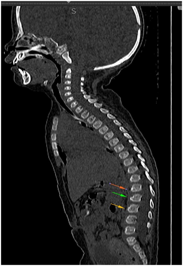
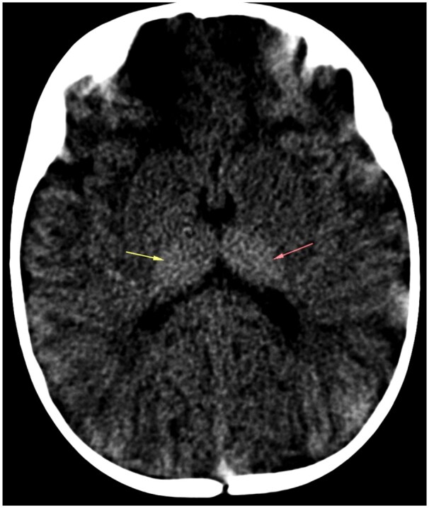
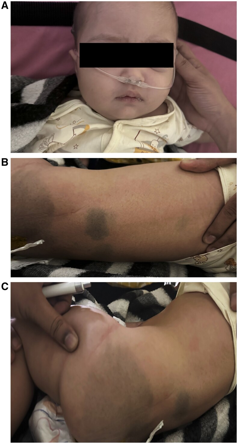
Mono-sialo-tetra-hexosylganglioside, also known as infantile GM1 gangliosidosis, is an autosomal recessive lysosomal storage disorder caused by a mutation in the GLB1 gene that stops the β-galactosidase enzyme from working. We have discussed a case of infantile GM1 gangliosidosis which presented with abnormal body movements, extensive dermal melanocytosis over back and gluteal region, coarse facial features, and macrocephaly. Radiological features included antero-inferior beaking of second, third, and fourth lumbar vertebrae, bilateral hyperdense thalami on non-contrast CT. On T2-weighted images, there is a persistently high signal intensity of the white matter and subcortical U fibres, which indicates bilateral bulky thalami with T2 hypointense and significantly impaired myelination. Reduced β-galactosidase activity verified the diagnosis.

 求助内容:
求助内容: 应助结果提醒方式:


