{"title":"副乳腺癌前哨淋巴结活检成功。","authors":"Young Duck Shin, Young Jin Choi","doi":"10.14740/jmc5094","DOIUrl":null,"url":null,"abstract":"<p><p>Primary breast cancer occurring in accessory breast tissue is exceptionally rare, with an incidence of 0.2-0.6%. It can aggressively progress, often leading to early metastasis. Treatment is typically delayed due to the rarity, variety of differentials, and lack of clinical awareness of the disease. In axillary surgery, sentinel lymph node mapping in patients with axillary breast cancer is technically challenging and has been poorly described. Here, we present a case of a 53-year-old woman with a 0.5 × 1 cm hard lump in the right axillary region for 2 years, progressive growth for 6 months, and no concomitant breast lesion or axillary lymphadenopathy. Core needle biopsy revealed invasive ductal carcinoma with estrogen receptor and progesterone receptor expression and human epidermal growth factor receptor 2 negativity, whereas mammography and breast magnetic resonance imaging revealed no primary breast lesions. She was diagnosed with invasive cancer arising from an accessory breast and underwent wide total excision of the right accessory breast and sentinel lymph node biopsy. Sentinel lymph node biopsy can be successfully performed using intratumoral dye and subareolar radiocolloid mapping in accessory breast cancer surgery. Axillary accessory breast tissue is outside the scope of the screening breast examination; therefore, oncologists must be aware of this entity and associated pathologies.</p>","PeriodicalId":101328,"journal":{"name":"Journal of medical cases","volume":"16 3","pages":"107-113"},"PeriodicalIF":0.9000,"publicationDate":"2025-03-01","publicationTypes":"Journal Article","fieldsOfStudy":null,"isOpenAccess":false,"openAccessPdf":"https://www.ncbi.nlm.nih.gov/pmc/articles/PMC11954603/pdf/","citationCount":"0","resultStr":"{\"title\":\"Successful Sentinel Lymph Node Biopsy in Accessory Breast Cancer.\",\"authors\":\"Young Duck Shin, Young Jin Choi\",\"doi\":\"10.14740/jmc5094\",\"DOIUrl\":null,\"url\":null,\"abstract\":\"<p><p>Primary breast cancer occurring in accessory breast tissue is exceptionally rare, with an incidence of 0.2-0.6%. It can aggressively progress, often leading to early metastasis. Treatment is typically delayed due to the rarity, variety of differentials, and lack of clinical awareness of the disease. In axillary surgery, sentinel lymph node mapping in patients with axillary breast cancer is technically challenging and has been poorly described. Here, we present a case of a 53-year-old woman with a 0.5 × 1 cm hard lump in the right axillary region for 2 years, progressive growth for 6 months, and no concomitant breast lesion or axillary lymphadenopathy. Core needle biopsy revealed invasive ductal carcinoma with estrogen receptor and progesterone receptor expression and human epidermal growth factor receptor 2 negativity, whereas mammography and breast magnetic resonance imaging revealed no primary breast lesions. She was diagnosed with invasive cancer arising from an accessory breast and underwent wide total excision of the right accessory breast and sentinel lymph node biopsy. Sentinel lymph node biopsy can be successfully performed using intratumoral dye and subareolar radiocolloid mapping in accessory breast cancer surgery. Axillary accessory breast tissue is outside the scope of the screening breast examination; therefore, oncologists must be aware of this entity and associated pathologies.</p>\",\"PeriodicalId\":101328,\"journal\":{\"name\":\"Journal of medical cases\",\"volume\":\"16 3\",\"pages\":\"107-113\"},\"PeriodicalIF\":0.9000,\"publicationDate\":\"2025-03-01\",\"publicationTypes\":\"Journal Article\",\"fieldsOfStudy\":null,\"isOpenAccess\":false,\"openAccessPdf\":\"https://www.ncbi.nlm.nih.gov/pmc/articles/PMC11954603/pdf/\",\"citationCount\":\"0\",\"resultStr\":null,\"platform\":\"Semanticscholar\",\"paperid\":null,\"PeriodicalName\":\"Journal of medical cases\",\"FirstCategoryId\":\"1085\",\"ListUrlMain\":\"https://doi.org/10.14740/jmc5094\",\"RegionNum\":0,\"RegionCategory\":null,\"ArticlePicture\":[],\"TitleCN\":null,\"AbstractTextCN\":null,\"PMCID\":null,\"EPubDate\":\"2025/2/18 0:00:00\",\"PubModel\":\"Epub\",\"JCR\":\"\",\"JCRName\":\"\",\"Score\":null,\"Total\":0}","platform":"Semanticscholar","paperid":null,"PeriodicalName":"Journal of medical cases","FirstCategoryId":"1085","ListUrlMain":"https://doi.org/10.14740/jmc5094","RegionNum":0,"RegionCategory":null,"ArticlePicture":[],"TitleCN":null,"AbstractTextCN":null,"PMCID":null,"EPubDate":"2025/2/18 0:00:00","PubModel":"Epub","JCR":"","JCRName":"","Score":null,"Total":0}
Successful Sentinel Lymph Node Biopsy in Accessory Breast Cancer.
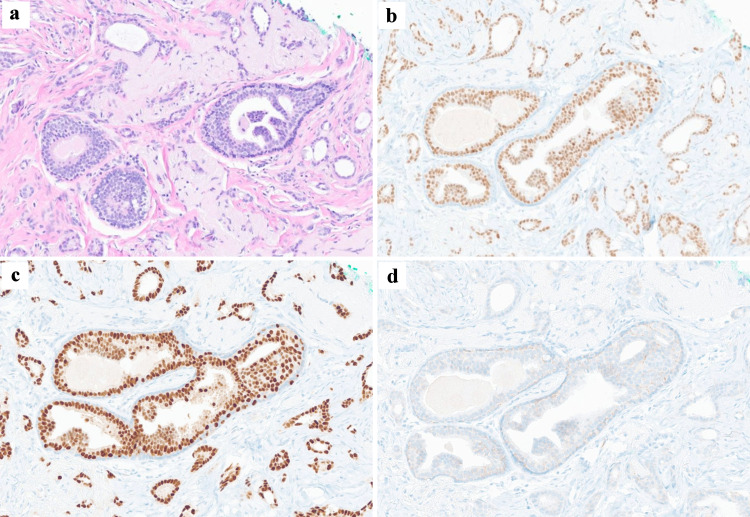
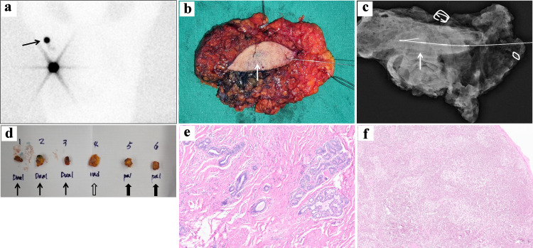
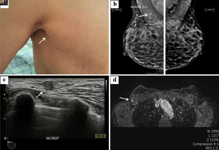
Primary breast cancer occurring in accessory breast tissue is exceptionally rare, with an incidence of 0.2-0.6%. It can aggressively progress, often leading to early metastasis. Treatment is typically delayed due to the rarity, variety of differentials, and lack of clinical awareness of the disease. In axillary surgery, sentinel lymph node mapping in patients with axillary breast cancer is technically challenging and has been poorly described. Here, we present a case of a 53-year-old woman with a 0.5 × 1 cm hard lump in the right axillary region for 2 years, progressive growth for 6 months, and no concomitant breast lesion or axillary lymphadenopathy. Core needle biopsy revealed invasive ductal carcinoma with estrogen receptor and progesterone receptor expression and human epidermal growth factor receptor 2 negativity, whereas mammography and breast magnetic resonance imaging revealed no primary breast lesions. She was diagnosed with invasive cancer arising from an accessory breast and underwent wide total excision of the right accessory breast and sentinel lymph node biopsy. Sentinel lymph node biopsy can be successfully performed using intratumoral dye and subareolar radiocolloid mapping in accessory breast cancer surgery. Axillary accessory breast tissue is outside the scope of the screening breast examination; therefore, oncologists must be aware of this entity and associated pathologies.

 求助内容:
求助内容: 应助结果提醒方式:


