Giorgio Dolci, Charles A Ellis, Federica Cruciani, Lorenza Brusini, Anees Abrol, Ilaria Boscolo Galazzo, Gloria Menegaz, Vince D Calhoun
{"title":"多模态MRI准确识别阿尔茨海默病连续体中不平衡队列中的淀粉样蛋白状态。","authors":"Giorgio Dolci, Charles A Ellis, Federica Cruciani, Lorenza Brusini, Anees Abrol, Ilaria Boscolo Galazzo, Gloria Menegaz, Vince D Calhoun","doi":"10.1162/netn_a_00423","DOIUrl":null,"url":null,"abstract":"<p><p>Amyloid-<i>β</i> (A<i>β</i>) plaques in conjunction with hyperphosphorylated tau proteins in the form of neurofibrillary tangles are the two neuropathological hallmarks of Alzheimer's disease. It is well-known that the identification of individuals with A<i>β</i> positivity could enable early diagnosis. In this work, we aim at capturing the A<i>β</i> positivity status in an unbalanced cohort enclosing subjects at different disease stages, exploiting the underlying structural and connectivity disease-induced modulations as revealed by structural, functional, and diffusion MRI. Of note, due to the unbalanced cohort, the outcomes may be guided by those factors rather than amyloid accumulation. The partial views provided by each modality are integrated in the model, allowing to take full advantage of their complementarity in encoding the effects of the A<i>β</i> accumulation, leading to an accuracy of 0.762 ± 0.04. The specificity of the information brought by each modality is assessed by post hoc explainability analysis (guided backpropagation), highlighting the underlying structural and functional changes. Noteworthy, well-established biomarker key regions related to A<i>β</i> deposition could be identified by all modalities, including the hippocampus, thalamus, precuneus, and cingulate gyrus, witnessing in favor of the reliability of the method as well as its potential in shedding light on modality-specific possibly unknown A<i>β</i> deposition signatures.</p>","PeriodicalId":48520,"journal":{"name":"Network Neuroscience","volume":"9 1","pages":"259-279"},"PeriodicalIF":3.1000,"publicationDate":"2025-03-20","publicationTypes":"Journal Article","fieldsOfStudy":null,"isOpenAccess":false,"openAccessPdf":"https://www.ncbi.nlm.nih.gov/pmc/articles/PMC11949592/pdf/","citationCount":"0","resultStr":"{\"title\":\"Multimodal MRI accurately identifies amyloid status in unbalanced cohorts in Alzheimer's disease continuum.\",\"authors\":\"Giorgio Dolci, Charles A Ellis, Federica Cruciani, Lorenza Brusini, Anees Abrol, Ilaria Boscolo Galazzo, Gloria Menegaz, Vince D Calhoun\",\"doi\":\"10.1162/netn_a_00423\",\"DOIUrl\":null,\"url\":null,\"abstract\":\"<p><p>Amyloid-<i>β</i> (A<i>β</i>) plaques in conjunction with hyperphosphorylated tau proteins in the form of neurofibrillary tangles are the two neuropathological hallmarks of Alzheimer's disease. It is well-known that the identification of individuals with A<i>β</i> positivity could enable early diagnosis. In this work, we aim at capturing the A<i>β</i> positivity status in an unbalanced cohort enclosing subjects at different disease stages, exploiting the underlying structural and connectivity disease-induced modulations as revealed by structural, functional, and diffusion MRI. Of note, due to the unbalanced cohort, the outcomes may be guided by those factors rather than amyloid accumulation. The partial views provided by each modality are integrated in the model, allowing to take full advantage of their complementarity in encoding the effects of the A<i>β</i> accumulation, leading to an accuracy of 0.762 ± 0.04. The specificity of the information brought by each modality is assessed by post hoc explainability analysis (guided backpropagation), highlighting the underlying structural and functional changes. Noteworthy, well-established biomarker key regions related to A<i>β</i> deposition could be identified by all modalities, including the hippocampus, thalamus, precuneus, and cingulate gyrus, witnessing in favor of the reliability of the method as well as its potential in shedding light on modality-specific possibly unknown A<i>β</i> deposition signatures.</p>\",\"PeriodicalId\":48520,\"journal\":{\"name\":\"Network Neuroscience\",\"volume\":\"9 1\",\"pages\":\"259-279\"},\"PeriodicalIF\":3.1000,\"publicationDate\":\"2025-03-20\",\"publicationTypes\":\"Journal Article\",\"fieldsOfStudy\":null,\"isOpenAccess\":false,\"openAccessPdf\":\"https://www.ncbi.nlm.nih.gov/pmc/articles/PMC11949592/pdf/\",\"citationCount\":\"0\",\"resultStr\":null,\"platform\":\"Semanticscholar\",\"paperid\":null,\"PeriodicalName\":\"Network Neuroscience\",\"FirstCategoryId\":\"3\",\"ListUrlMain\":\"https://doi.org/10.1162/netn_a_00423\",\"RegionNum\":3,\"RegionCategory\":\"医学\",\"ArticlePicture\":[],\"TitleCN\":null,\"AbstractTextCN\":null,\"PMCID\":null,\"EPubDate\":\"2025/1/1 0:00:00\",\"PubModel\":\"eCollection\",\"JCR\":\"Q2\",\"JCRName\":\"NEUROSCIENCES\",\"Score\":null,\"Total\":0}","platform":"Semanticscholar","paperid":null,"PeriodicalName":"Network Neuroscience","FirstCategoryId":"3","ListUrlMain":"https://doi.org/10.1162/netn_a_00423","RegionNum":3,"RegionCategory":"医学","ArticlePicture":[],"TitleCN":null,"AbstractTextCN":null,"PMCID":null,"EPubDate":"2025/1/1 0:00:00","PubModel":"eCollection","JCR":"Q2","JCRName":"NEUROSCIENCES","Score":null,"Total":0}
Multimodal MRI accurately identifies amyloid status in unbalanced cohorts in Alzheimer's disease continuum.
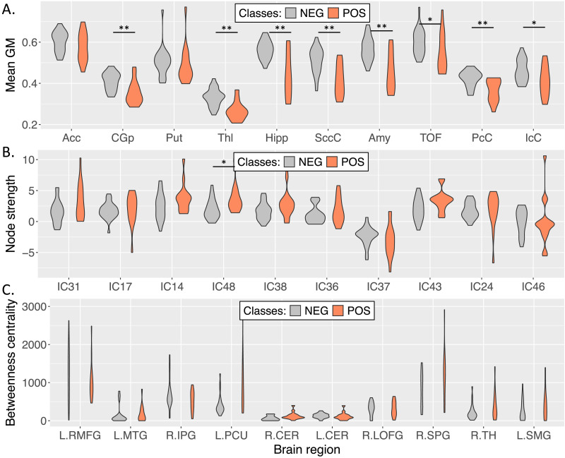
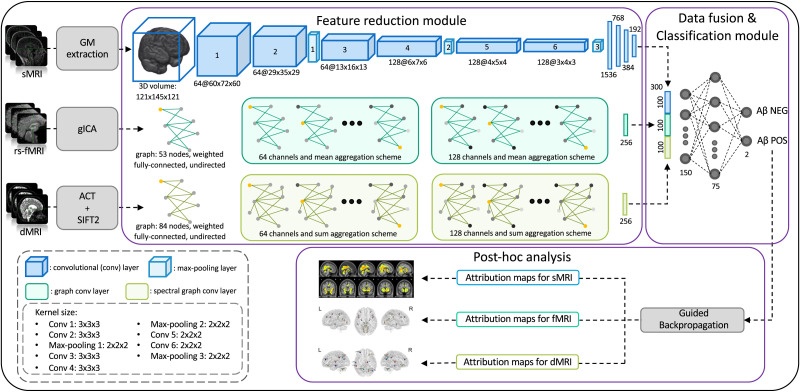
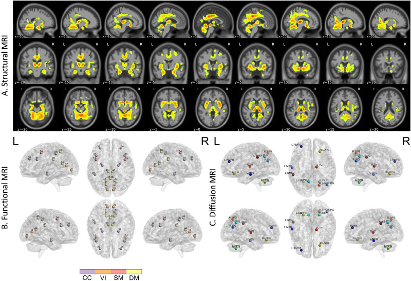
Amyloid-β (Aβ) plaques in conjunction with hyperphosphorylated tau proteins in the form of neurofibrillary tangles are the two neuropathological hallmarks of Alzheimer's disease. It is well-known that the identification of individuals with Aβ positivity could enable early diagnosis. In this work, we aim at capturing the Aβ positivity status in an unbalanced cohort enclosing subjects at different disease stages, exploiting the underlying structural and connectivity disease-induced modulations as revealed by structural, functional, and diffusion MRI. Of note, due to the unbalanced cohort, the outcomes may be guided by those factors rather than amyloid accumulation. The partial views provided by each modality are integrated in the model, allowing to take full advantage of their complementarity in encoding the effects of the Aβ accumulation, leading to an accuracy of 0.762 ± 0.04. The specificity of the information brought by each modality is assessed by post hoc explainability analysis (guided backpropagation), highlighting the underlying structural and functional changes. Noteworthy, well-established biomarker key regions related to Aβ deposition could be identified by all modalities, including the hippocampus, thalamus, precuneus, and cingulate gyrus, witnessing in favor of the reliability of the method as well as its potential in shedding light on modality-specific possibly unknown Aβ deposition signatures.
 求助内容:
求助内容: 应助结果提醒方式:


