Xinyuan Zheng, Patrick Worhunsky, Qiong Liu, Xueqi Guo, Xiongchao Chen, Heng Sun, Jiazhen Zhang, Takuya Toyonaga, Adam P Mecca, Ryan S O'Dell, Christopher H van Dyck, Gustavo A Angarita, Kelly Cosgrove, Deepak D'Souza, David Matuskey, Irina Esterlis, Richard E Carson, Rajiv Radhakrishnan, Chi Liu
{"title":"基于磁共振T1图像,利用深度学习生成突触密度的合成脑PET图像。","authors":"Xinyuan Zheng, Patrick Worhunsky, Qiong Liu, Xueqi Guo, Xiongchao Chen, Heng Sun, Jiazhen Zhang, Takuya Toyonaga, Adam P Mecca, Ryan S O'Dell, Christopher H van Dyck, Gustavo A Angarita, Kelly Cosgrove, Deepak D'Souza, David Matuskey, Irina Esterlis, Richard E Carson, Rajiv Radhakrishnan, Chi Liu","doi":"10.1186/s40658-025-00744-5","DOIUrl":null,"url":null,"abstract":"<p><strong>Purpose: </strong>Synaptic vesicle glycoprotein 2 A (SV2A) in human brains is an important biomarker of synaptic loss associated with several neurological disorders. However, SV2A tracers, such as [<sup>11</sup>C]UCB-J, are less available in practice due to constrains such as cost, radiation exposure and onsite cyclotron. We therefore aim to generate synthetic [<sup>11</sup>C]UCB-J PET images based on MRI in this study.</p><p><strong>Methods: </strong>We implemented a convolution-based 3D encoder-decoder to predict [<sup>11</sup>C]UCB-J SV2A PET images. A total of 160 participants who underwent both MRI and [<sup>11</sup>C]UCB-J PET imaging, including individuals with schizophrenia, cannabis use disorder, Alzheimer's disease, were used in this study. The model was trained on pairs of T1-weighted MRI and [<sup>11</sup>C]UCB-J distribution volume ratio images, and tested through a 10-fold cross-validation process. The image translation accuracy was evaluated based on the mean squared error, structural similarity index, percentage bias and Pearson's correlation coefficient between the ground truth and the predicted images. Additionally, we assessed the prediction accuracy of selected regions of interest (ROIs) crucial for brain disorders to evaluate our results.</p><p><strong>Results: </strong>The generated SV2A PET images are visually similar to the ground truth in terms of contrast and tracer distribution, quantitatively with low bias (< 2%) and high similarity (> 0.9). Across all diagnostic categories and ROIs, including the hippocampus, frontal, occipital, parietal, and temporal regions, the synthetic SV2A PET images exhibit an average bias of less than 5% compared to the ground truth. The model also demonstrates a capacity for noise reduction, producing images of higher quality compared to the low-dose scans.</p><p><strong>Conclusion: </strong>We conclude that it is feasible to generate robust SV2A PET images with promising accuracy from MRI via a data-driven approach.</p>","PeriodicalId":11559,"journal":{"name":"EJNMMI Physics","volume":"12 1","pages":"30"},"PeriodicalIF":3.2000,"publicationDate":"2025-03-31","publicationTypes":"Journal Article","fieldsOfStudy":null,"isOpenAccess":false,"openAccessPdf":"https://www.ncbi.nlm.nih.gov/pmc/articles/PMC11958861/pdf/","citationCount":"0","resultStr":"{\"title\":\"Generating synthetic brain PET images of synaptic density based on MR T1 images using deep learning.\",\"authors\":\"Xinyuan Zheng, Patrick Worhunsky, Qiong Liu, Xueqi Guo, Xiongchao Chen, Heng Sun, Jiazhen Zhang, Takuya Toyonaga, Adam P Mecca, Ryan S O'Dell, Christopher H van Dyck, Gustavo A Angarita, Kelly Cosgrove, Deepak D'Souza, David Matuskey, Irina Esterlis, Richard E Carson, Rajiv Radhakrishnan, Chi Liu\",\"doi\":\"10.1186/s40658-025-00744-5\",\"DOIUrl\":null,\"url\":null,\"abstract\":\"<p><strong>Purpose: </strong>Synaptic vesicle glycoprotein 2 A (SV2A) in human brains is an important biomarker of synaptic loss associated with several neurological disorders. However, SV2A tracers, such as [<sup>11</sup>C]UCB-J, are less available in practice due to constrains such as cost, radiation exposure and onsite cyclotron. We therefore aim to generate synthetic [<sup>11</sup>C]UCB-J PET images based on MRI in this study.</p><p><strong>Methods: </strong>We implemented a convolution-based 3D encoder-decoder to predict [<sup>11</sup>C]UCB-J SV2A PET images. A total of 160 participants who underwent both MRI and [<sup>11</sup>C]UCB-J PET imaging, including individuals with schizophrenia, cannabis use disorder, Alzheimer's disease, were used in this study. The model was trained on pairs of T1-weighted MRI and [<sup>11</sup>C]UCB-J distribution volume ratio images, and tested through a 10-fold cross-validation process. The image translation accuracy was evaluated based on the mean squared error, structural similarity index, percentage bias and Pearson's correlation coefficient between the ground truth and the predicted images. Additionally, we assessed the prediction accuracy of selected regions of interest (ROIs) crucial for brain disorders to evaluate our results.</p><p><strong>Results: </strong>The generated SV2A PET images are visually similar to the ground truth in terms of contrast and tracer distribution, quantitatively with low bias (< 2%) and high similarity (> 0.9). Across all diagnostic categories and ROIs, including the hippocampus, frontal, occipital, parietal, and temporal regions, the synthetic SV2A PET images exhibit an average bias of less than 5% compared to the ground truth. The model also demonstrates a capacity for noise reduction, producing images of higher quality compared to the low-dose scans.</p><p><strong>Conclusion: </strong>We conclude that it is feasible to generate robust SV2A PET images with promising accuracy from MRI via a data-driven approach.</p>\",\"PeriodicalId\":11559,\"journal\":{\"name\":\"EJNMMI Physics\",\"volume\":\"12 1\",\"pages\":\"30\"},\"PeriodicalIF\":3.2000,\"publicationDate\":\"2025-03-31\",\"publicationTypes\":\"Journal Article\",\"fieldsOfStudy\":null,\"isOpenAccess\":false,\"openAccessPdf\":\"https://www.ncbi.nlm.nih.gov/pmc/articles/PMC11958861/pdf/\",\"citationCount\":\"0\",\"resultStr\":null,\"platform\":\"Semanticscholar\",\"paperid\":null,\"PeriodicalName\":\"EJNMMI Physics\",\"FirstCategoryId\":\"3\",\"ListUrlMain\":\"https://doi.org/10.1186/s40658-025-00744-5\",\"RegionNum\":2,\"RegionCategory\":\"医学\",\"ArticlePicture\":[],\"TitleCN\":null,\"AbstractTextCN\":null,\"PMCID\":null,\"EPubDate\":\"\",\"PubModel\":\"\",\"JCR\":\"Q2\",\"JCRName\":\"RADIOLOGY, NUCLEAR MEDICINE & MEDICAL IMAGING\",\"Score\":null,\"Total\":0}","platform":"Semanticscholar","paperid":null,"PeriodicalName":"EJNMMI Physics","FirstCategoryId":"3","ListUrlMain":"https://doi.org/10.1186/s40658-025-00744-5","RegionNum":2,"RegionCategory":"医学","ArticlePicture":[],"TitleCN":null,"AbstractTextCN":null,"PMCID":null,"EPubDate":"","PubModel":"","JCR":"Q2","JCRName":"RADIOLOGY, NUCLEAR MEDICINE & MEDICAL IMAGING","Score":null,"Total":0}
引用次数: 0
摘要
目的:人脑中的突触小泡糖蛋白 2 A(SV2A)是与多种神经系统疾病相关的突触损失的重要生物标志物。然而,由于受到成本、辐射暴露和现场回旋加速器等因素的限制,[11C]UCB-J 等 SV2A 示踪剂在实际应用中较少。因此,本研究旨在基于核磁共振成像生成合成的 [11C]UCB-J PET 图像:我们采用基于卷积的三维编码器-解码器来预测[11C]UCB-J SV2A PET图像。本研究共使用了 160 名同时接受 MRI 和 [11C]UCB-J PET 成像检查的参与者,其中包括精神分裂症患者、大麻使用障碍患者和阿尔茨海默病患者。该模型在成对的 T1 加权 MRI 和 [11C]UCB-J 分布容积比图像上进行训练,并通过 10 倍交叉验证过程进行测试。根据地面实况和预测图像之间的均方误差、结构相似性指数、偏差百分比和皮尔逊相关系数评估了图像转换的准确性。此外,我们还评估了对脑部疾病至关重要的选定感兴趣区(ROI)的预测准确性,以评价我们的结果:结果:在对比度和示踪剂分布方面,生成的 SV2A PET 图像在视觉上与地面实况相似,在数量上偏差较小(0.9)。在包括海马、额叶、枕叶、顶叶和颞叶区域在内的所有诊断类别和 ROI 中,合成 SV2A PET 图像与地面实况相比平均偏差小于 5%。该模型还具有降噪能力,生成的图像质量高于低剂量扫描图像:我们的结论是,通过数据驱动方法从核磁共振成像生成具有良好准确性的稳健 SV2A PET 图像是可行的。
Generating synthetic brain PET images of synaptic density based on MR T1 images using deep learning.
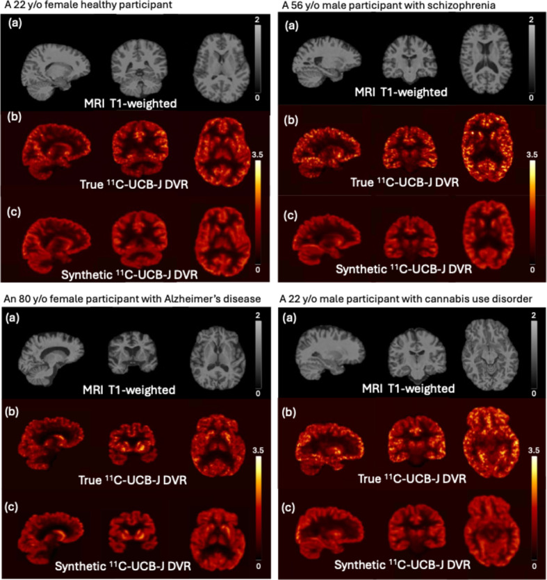
Purpose: Synaptic vesicle glycoprotein 2 A (SV2A) in human brains is an important biomarker of synaptic loss associated with several neurological disorders. However, SV2A tracers, such as [11C]UCB-J, are less available in practice due to constrains such as cost, radiation exposure and onsite cyclotron. We therefore aim to generate synthetic [11C]UCB-J PET images based on MRI in this study.
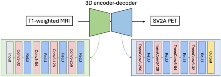
Methods: We implemented a convolution-based 3D encoder-decoder to predict [11C]UCB-J SV2A PET images. A total of 160 participants who underwent both MRI and [11C]UCB-J PET imaging, including individuals with schizophrenia, cannabis use disorder, Alzheimer's disease, were used in this study. The model was trained on pairs of T1-weighted MRI and [11C]UCB-J distribution volume ratio images, and tested through a 10-fold cross-validation process. The image translation accuracy was evaluated based on the mean squared error, structural similarity index, percentage bias and Pearson's correlation coefficient between the ground truth and the predicted images. Additionally, we assessed the prediction accuracy of selected regions of interest (ROIs) crucial for brain disorders to evaluate our results.
Results: The generated SV2A PET images are visually similar to the ground truth in terms of contrast and tracer distribution, quantitatively with low bias (< 2%) and high similarity (> 0.9). Across all diagnostic categories and ROIs, including the hippocampus, frontal, occipital, parietal, and temporal regions, the synthetic SV2A PET images exhibit an average bias of less than 5% compared to the ground truth. The model also demonstrates a capacity for noise reduction, producing images of higher quality compared to the low-dose scans.
Conclusion: We conclude that it is feasible to generate robust SV2A PET images with promising accuracy from MRI via a data-driven approach.
期刊介绍:
EJNMMI Physics is an international platform for scientists, users and adopters of nuclear medicine with a particular interest in physics matters. As a companion journal to the European Journal of Nuclear Medicine and Molecular Imaging, this journal has a multi-disciplinary approach and welcomes original materials and studies with a focus on applied physics and mathematics as well as imaging systems engineering and prototyping in nuclear medicine. This includes physics-driven approaches or algorithms supported by physics that foster early clinical adoption of nuclear medicine imaging and therapy.

 求助内容:
求助内容: 应助结果提醒方式:


