{"title":"基于虚拟现实的介入放射学模拟器在医学生教育中的有效性。","authors":"Hidenori Mitani, Yukiko Honda, Keigo Narita, Yuko Nakamura, Shintaro Morishita, Shota Kondo, Shogo Maeda, Haruka Higashibori, Keigo Chosa, Toru Higaki, Ikuo Kawashita, Minoru Hattori, Naoko Hasunuma, Isamu Saeki, Shinya Takahashi, Naoki Mihara, Kazuo Awai","doi":"10.1007/s11604-025-01771-z","DOIUrl":null,"url":null,"abstract":"<p><strong>Purpose: </strong>We developed an interventional radiology (IR) simulator using a virtual reality system (the VR-IR simulator) to teach IR procedures to medical students. In this study, we investigated the effectiveness of this teaching method.</p><p><strong>Materials and methods: </strong>All ninety-nine fifth-year medical students attended a conventional classroom lecture. To teach students the actual procedure, they were randomly divided into two groups: One received conventional verbal explanations and educator demonstrations (the conventional group [n = 44]), and the other received VR-IR simulator training (the VR-IR simulator group [n = 55]). Afterward, they underwent a test using an augmented reality- (AR-) IR simulator (the VIST<sup>®</sup> G5 image-guided AR-IR simulator, Mentice, Gothenburg, Sweden). The total procedure time, amount of contrast media used, fluoroscopic time, and patient peak skin dose in the simulated patients were compared between groups. A board-certified radiologist evaluated ten aspects of the procedure technique using a 5-point Likert scale (total: 50 points).</p><p><strong>Results: </strong>Two students in the VR-IR simulator group were excluded due to VR sickness and simulator malfunction. There were no significant differences between the VR-IR simulator group and the conventional group regarding total procedure time (median [25-75% interquartile range]: 13.5 [11.8-14.5] vs. 14.3 [12.3-16.8] minutes, p = 0.11), fluoroscopic time (10.1 [8.5-13.0] vs. 11.0 [8.6-13.7] minutes, p = 0.31), and patient peak skin dose (276 [243-373] vs. 303 [239-395] mGy, p = 0.57), respectively. However, the amount of contrast media used was significantly lower (28.0 [21.0-36.2] vs. 40.0 [32.3-50.9] mL, p < 0.01) and the technical achievement scores by the radiologist (36 [34-44] vs. 31 [29-32], p < 0.01) were significantly higher in the VR-IR simulator group.</p><p><strong>Conclusion: </strong>The VR-IR simulator helped reduce the amount of contrast media in interventional procedures and improved technical achievement scores.</p>","PeriodicalId":14691,"journal":{"name":"Japanese Journal of Radiology","volume":" ","pages":"1386-1392"},"PeriodicalIF":2.1000,"publicationDate":"2025-08-01","publicationTypes":"Journal Article","fieldsOfStudy":null,"isOpenAccess":false,"openAccessPdf":"https://www.ncbi.nlm.nih.gov/pmc/articles/PMC12287170/pdf/","citationCount":"0","resultStr":"{\"title\":\"Effectiveness of a virtual reality-based interventional radiology simulator for medical student education.\",\"authors\":\"Hidenori Mitani, Yukiko Honda, Keigo Narita, Yuko Nakamura, Shintaro Morishita, Shota Kondo, Shogo Maeda, Haruka Higashibori, Keigo Chosa, Toru Higaki, Ikuo Kawashita, Minoru Hattori, Naoko Hasunuma, Isamu Saeki, Shinya Takahashi, Naoki Mihara, Kazuo Awai\",\"doi\":\"10.1007/s11604-025-01771-z\",\"DOIUrl\":null,\"url\":null,\"abstract\":\"<p><strong>Purpose: </strong>We developed an interventional radiology (IR) simulator using a virtual reality system (the VR-IR simulator) to teach IR procedures to medical students. In this study, we investigated the effectiveness of this teaching method.</p><p><strong>Materials and methods: </strong>All ninety-nine fifth-year medical students attended a conventional classroom lecture. To teach students the actual procedure, they were randomly divided into two groups: One received conventional verbal explanations and educator demonstrations (the conventional group [n = 44]), and the other received VR-IR simulator training (the VR-IR simulator group [n = 55]). Afterward, they underwent a test using an augmented reality- (AR-) IR simulator (the VIST<sup>®</sup> G5 image-guided AR-IR simulator, Mentice, Gothenburg, Sweden). The total procedure time, amount of contrast media used, fluoroscopic time, and patient peak skin dose in the simulated patients were compared between groups. A board-certified radiologist evaluated ten aspects of the procedure technique using a 5-point Likert scale (total: 50 points).</p><p><strong>Results: </strong>Two students in the VR-IR simulator group were excluded due to VR sickness and simulator malfunction. There were no significant differences between the VR-IR simulator group and the conventional group regarding total procedure time (median [25-75% interquartile range]: 13.5 [11.8-14.5] vs. 14.3 [12.3-16.8] minutes, p = 0.11), fluoroscopic time (10.1 [8.5-13.0] vs. 11.0 [8.6-13.7] minutes, p = 0.31), and patient peak skin dose (276 [243-373] vs. 303 [239-395] mGy, p = 0.57), respectively. However, the amount of contrast media used was significantly lower (28.0 [21.0-36.2] vs. 40.0 [32.3-50.9] mL, p < 0.01) and the technical achievement scores by the radiologist (36 [34-44] vs. 31 [29-32], p < 0.01) were significantly higher in the VR-IR simulator group.</p><p><strong>Conclusion: </strong>The VR-IR simulator helped reduce the amount of contrast media in interventional procedures and improved technical achievement scores.</p>\",\"PeriodicalId\":14691,\"journal\":{\"name\":\"Japanese Journal of Radiology\",\"volume\":\" \",\"pages\":\"1386-1392\"},\"PeriodicalIF\":2.1000,\"publicationDate\":\"2025-08-01\",\"publicationTypes\":\"Journal Article\",\"fieldsOfStudy\":null,\"isOpenAccess\":false,\"openAccessPdf\":\"https://www.ncbi.nlm.nih.gov/pmc/articles/PMC12287170/pdf/\",\"citationCount\":\"0\",\"resultStr\":null,\"platform\":\"Semanticscholar\",\"paperid\":null,\"PeriodicalName\":\"Japanese Journal of Radiology\",\"FirstCategoryId\":\"3\",\"ListUrlMain\":\"https://doi.org/10.1007/s11604-025-01771-z\",\"RegionNum\":4,\"RegionCategory\":\"医学\",\"ArticlePicture\":[],\"TitleCN\":null,\"AbstractTextCN\":null,\"PMCID\":null,\"EPubDate\":\"2025/3/29 0:00:00\",\"PubModel\":\"Epub\",\"JCR\":\"\",\"JCRName\":\"\",\"Score\":null,\"Total\":0}","platform":"Semanticscholar","paperid":null,"PeriodicalName":"Japanese Journal of Radiology","FirstCategoryId":"3","ListUrlMain":"https://doi.org/10.1007/s11604-025-01771-z","RegionNum":4,"RegionCategory":"医学","ArticlePicture":[],"TitleCN":null,"AbstractTextCN":null,"PMCID":null,"EPubDate":"2025/3/29 0:00:00","PubModel":"Epub","JCR":"","JCRName":"","Score":null,"Total":0}
Effectiveness of a virtual reality-based interventional radiology simulator for medical student education.
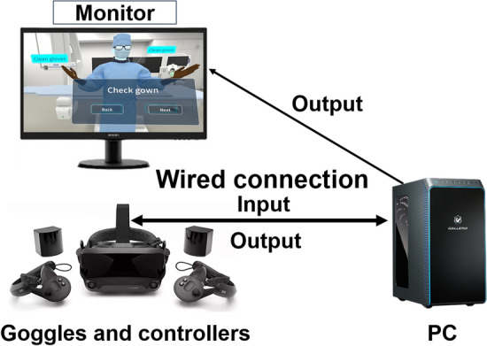
Purpose: We developed an interventional radiology (IR) simulator using a virtual reality system (the VR-IR simulator) to teach IR procedures to medical students. In this study, we investigated the effectiveness of this teaching method.
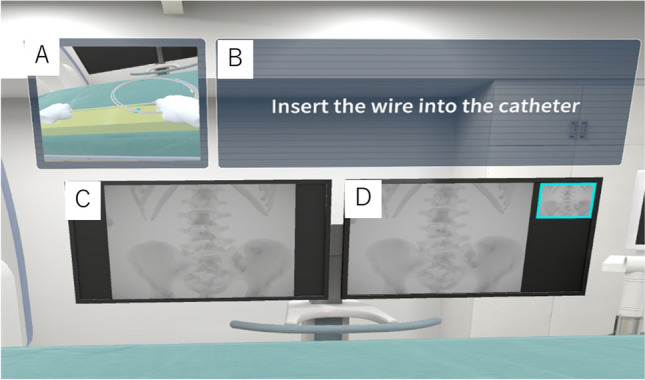
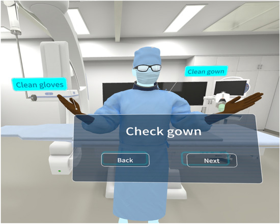
Materials and methods: All ninety-nine fifth-year medical students attended a conventional classroom lecture. To teach students the actual procedure, they were randomly divided into two groups: One received conventional verbal explanations and educator demonstrations (the conventional group [n = 44]), and the other received VR-IR simulator training (the VR-IR simulator group [n = 55]). Afterward, they underwent a test using an augmented reality- (AR-) IR simulator (the VIST® G5 image-guided AR-IR simulator, Mentice, Gothenburg, Sweden). The total procedure time, amount of contrast media used, fluoroscopic time, and patient peak skin dose in the simulated patients were compared between groups. A board-certified radiologist evaluated ten aspects of the procedure technique using a 5-point Likert scale (total: 50 points).
Results: Two students in the VR-IR simulator group were excluded due to VR sickness and simulator malfunction. There were no significant differences between the VR-IR simulator group and the conventional group regarding total procedure time (median [25-75% interquartile range]: 13.5 [11.8-14.5] vs. 14.3 [12.3-16.8] minutes, p = 0.11), fluoroscopic time (10.1 [8.5-13.0] vs. 11.0 [8.6-13.7] minutes, p = 0.31), and patient peak skin dose (276 [243-373] vs. 303 [239-395] mGy, p = 0.57), respectively. However, the amount of contrast media used was significantly lower (28.0 [21.0-36.2] vs. 40.0 [32.3-50.9] mL, p < 0.01) and the technical achievement scores by the radiologist (36 [34-44] vs. 31 [29-32], p < 0.01) were significantly higher in the VR-IR simulator group.
Conclusion: The VR-IR simulator helped reduce the amount of contrast media in interventional procedures and improved technical achievement scores.
期刊介绍:
Japanese Journal of Radiology is a peer-reviewed journal, officially published by the Japan Radiological Society. The main purpose of the journal is to provide a forum for the publication of papers documenting recent advances and new developments in the field of radiology in medicine and biology. The scope of Japanese Journal of Radiology encompasses but is not restricted to diagnostic radiology, interventional radiology, radiation oncology, nuclear medicine, radiation physics, and radiation biology. Additionally, the journal covers technical and industrial innovations. The journal welcomes original articles, technical notes, review articles, pictorial essays and letters to the editor. The journal also provides announcements from the boards and the committees of the society. Membership in the Japan Radiological Society is not a prerequisite for submission. Contributions are welcomed from all parts of the world.

 求助内容:
求助内容: 应助结果提醒方式:


