Wen Li, Natsuko Onishi, Jessica E Gibbs, Lisa J Wilmes, Nu N Le, Pouya Metanat, Elissa R Price, Bonnie N Joe, John Kornak, Christina Yau, Denise M Wolf, Mark Jesus M Magbanua, Barbara LeStage, Laura J van 't Veer, Angela M DeMichele, Laura J Esserman, Nola M Hylton
{"title":"基于mri的乳腺癌个体化新辅助治疗模型。","authors":"Wen Li, Natsuko Onishi, Jessica E Gibbs, Lisa J Wilmes, Nu N Le, Pouya Metanat, Elissa R Price, Bonnie N Joe, John Kornak, Christina Yau, Denise M Wolf, Mark Jesus M Magbanua, Barbara LeStage, Laura J van 't Veer, Angela M DeMichele, Laura J Esserman, Nola M Hylton","doi":"10.3390/tomography11030026","DOIUrl":null,"url":null,"abstract":"<p><strong>Background: </strong>Functional tumor volume (FTV), measured from dynamic contrast-enhanced MRI, is an imaging biomarker that can predict treatment response in breast cancer patients undergoing neoadjuvant chemotherapy (NAC). The FTV-based predictive model, combined with core biopsy, informed treatment decisions of recommending patients with excellent responses to proceed to surgery early in a large NAC clinical trial.</p><p><strong>Methods: </strong>In this retrospective study, we constructed models using FTV measurements. We analyzed performance tradeoffs when a probability threshold was used to identify excellent responders through the prediction of pathology complete response (pCR). Individual models were developed within cohorts defined by the hormone receptor and human epidermal growth factor receptor 2 (HR/HER2) subtype.</p><p><strong>Results: </strong>A total of 814 patients enrolled in the I-SPY 2 trial between 2010 and 2016 were included with a mean age of 49 years (range: 24 to 77). Among these patients, 289 (36%) achieved pCR. The area under the ROC curve (AUC) ranged from 0.68 to 0.74 for individual HR/HER2 subtypes. When probability thresholds were chosen based on minimum positive predictive value (PPV) levels of 50%, 70%, and 90%, the PPV-sensitivity tradeoff varied among subtypes. The highest sensitivities (100%, 87%, 45%) were found in the HR-/HER2+ sub-cohort for probability thresholds of 0, 0.62, and 0.72; followed by the triple-negative sub-cohort (98%, 52%, 4%) at thresholds of 0.13, 0.58, and 0.67; and HR+/HER2+ (78%, 16%, 8%) at thresholds of 0.34, 0.57, and 0.60. The lowest sensitivities (20%, 0%, 0%) occurred in the HR+/HER2- sub-cohort.</p><p><strong>Conclusions: </strong>Predictive models developed using imaging biomarkers, alongside clinically validated probability thresholds, can be incorporated into decision-making for precision oncology.</p>","PeriodicalId":51330,"journal":{"name":"Tomography","volume":"11 3","pages":""},"PeriodicalIF":2.2000,"publicationDate":"2025-02-27","publicationTypes":"Journal Article","fieldsOfStudy":null,"isOpenAccess":false,"openAccessPdf":"https://www.ncbi.nlm.nih.gov/pmc/articles/PMC11946387/pdf/","citationCount":"0","resultStr":"{\"title\":\"MRI-Based Model for Personalizing Neoadjuvant Treatment in Breast Cancer.\",\"authors\":\"Wen Li, Natsuko Onishi, Jessica E Gibbs, Lisa J Wilmes, Nu N Le, Pouya Metanat, Elissa R Price, Bonnie N Joe, John Kornak, Christina Yau, Denise M Wolf, Mark Jesus M Magbanua, Barbara LeStage, Laura J van 't Veer, Angela M DeMichele, Laura J Esserman, Nola M Hylton\",\"doi\":\"10.3390/tomography11030026\",\"DOIUrl\":null,\"url\":null,\"abstract\":\"<p><strong>Background: </strong>Functional tumor volume (FTV), measured from dynamic contrast-enhanced MRI, is an imaging biomarker that can predict treatment response in breast cancer patients undergoing neoadjuvant chemotherapy (NAC). The FTV-based predictive model, combined with core biopsy, informed treatment decisions of recommending patients with excellent responses to proceed to surgery early in a large NAC clinical trial.</p><p><strong>Methods: </strong>In this retrospective study, we constructed models using FTV measurements. We analyzed performance tradeoffs when a probability threshold was used to identify excellent responders through the prediction of pathology complete response (pCR). Individual models were developed within cohorts defined by the hormone receptor and human epidermal growth factor receptor 2 (HR/HER2) subtype.</p><p><strong>Results: </strong>A total of 814 patients enrolled in the I-SPY 2 trial between 2010 and 2016 were included with a mean age of 49 years (range: 24 to 77). Among these patients, 289 (36%) achieved pCR. The area under the ROC curve (AUC) ranged from 0.68 to 0.74 for individual HR/HER2 subtypes. When probability thresholds were chosen based on minimum positive predictive value (PPV) levels of 50%, 70%, and 90%, the PPV-sensitivity tradeoff varied among subtypes. The highest sensitivities (100%, 87%, 45%) were found in the HR-/HER2+ sub-cohort for probability thresholds of 0, 0.62, and 0.72; followed by the triple-negative sub-cohort (98%, 52%, 4%) at thresholds of 0.13, 0.58, and 0.67; and HR+/HER2+ (78%, 16%, 8%) at thresholds of 0.34, 0.57, and 0.60. The lowest sensitivities (20%, 0%, 0%) occurred in the HR+/HER2- sub-cohort.</p><p><strong>Conclusions: </strong>Predictive models developed using imaging biomarkers, alongside clinically validated probability thresholds, can be incorporated into decision-making for precision oncology.</p>\",\"PeriodicalId\":51330,\"journal\":{\"name\":\"Tomography\",\"volume\":\"11 3\",\"pages\":\"\"},\"PeriodicalIF\":2.2000,\"publicationDate\":\"2025-02-27\",\"publicationTypes\":\"Journal Article\",\"fieldsOfStudy\":null,\"isOpenAccess\":false,\"openAccessPdf\":\"https://www.ncbi.nlm.nih.gov/pmc/articles/PMC11946387/pdf/\",\"citationCount\":\"0\",\"resultStr\":null,\"platform\":\"Semanticscholar\",\"paperid\":null,\"PeriodicalName\":\"Tomography\",\"FirstCategoryId\":\"3\",\"ListUrlMain\":\"https://doi.org/10.3390/tomography11030026\",\"RegionNum\":4,\"RegionCategory\":\"医学\",\"ArticlePicture\":[],\"TitleCN\":null,\"AbstractTextCN\":null,\"PMCID\":null,\"EPubDate\":\"\",\"PubModel\":\"\",\"JCR\":\"Q2\",\"JCRName\":\"RADIOLOGY, NUCLEAR MEDICINE & MEDICAL IMAGING\",\"Score\":null,\"Total\":0}","platform":"Semanticscholar","paperid":null,"PeriodicalName":"Tomography","FirstCategoryId":"3","ListUrlMain":"https://doi.org/10.3390/tomography11030026","RegionNum":4,"RegionCategory":"医学","ArticlePicture":[],"TitleCN":null,"AbstractTextCN":null,"PMCID":null,"EPubDate":"","PubModel":"","JCR":"Q2","JCRName":"RADIOLOGY, NUCLEAR MEDICINE & MEDICAL IMAGING","Score":null,"Total":0}
MRI-Based Model for Personalizing Neoadjuvant Treatment in Breast Cancer.
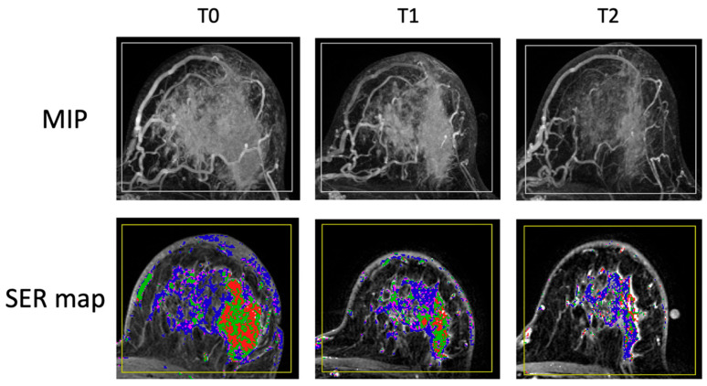
Background: Functional tumor volume (FTV), measured from dynamic contrast-enhanced MRI, is an imaging biomarker that can predict treatment response in breast cancer patients undergoing neoadjuvant chemotherapy (NAC). The FTV-based predictive model, combined with core biopsy, informed treatment decisions of recommending patients with excellent responses to proceed to surgery early in a large NAC clinical trial.
Methods: In this retrospective study, we constructed models using FTV measurements. We analyzed performance tradeoffs when a probability threshold was used to identify excellent responders through the prediction of pathology complete response (pCR). Individual models were developed within cohorts defined by the hormone receptor and human epidermal growth factor receptor 2 (HR/HER2) subtype.
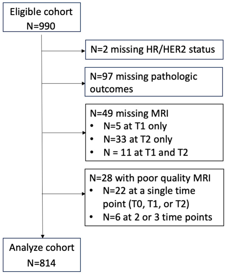
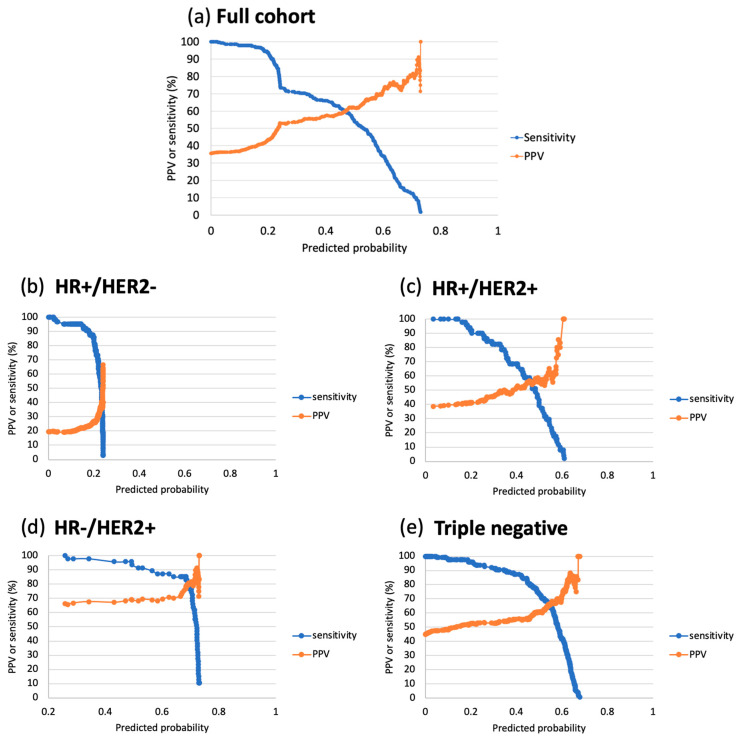
Results: A total of 814 patients enrolled in the I-SPY 2 trial between 2010 and 2016 were included with a mean age of 49 years (range: 24 to 77). Among these patients, 289 (36%) achieved pCR. The area under the ROC curve (AUC) ranged from 0.68 to 0.74 for individual HR/HER2 subtypes. When probability thresholds were chosen based on minimum positive predictive value (PPV) levels of 50%, 70%, and 90%, the PPV-sensitivity tradeoff varied among subtypes. The highest sensitivities (100%, 87%, 45%) were found in the HR-/HER2+ sub-cohort for probability thresholds of 0, 0.62, and 0.72; followed by the triple-negative sub-cohort (98%, 52%, 4%) at thresholds of 0.13, 0.58, and 0.67; and HR+/HER2+ (78%, 16%, 8%) at thresholds of 0.34, 0.57, and 0.60. The lowest sensitivities (20%, 0%, 0%) occurred in the HR+/HER2- sub-cohort.
Conclusions: Predictive models developed using imaging biomarkers, alongside clinically validated probability thresholds, can be incorporated into decision-making for precision oncology.
TomographyMedicine-Radiology, Nuclear Medicine and Imaging
CiteScore
2.70
自引率
10.50%
发文量
222
期刊介绍:
TomographyTM publishes basic (technical and pre-clinical) and clinical scientific articles which involve the advancement of imaging technologies. Tomography encompasses studies that use single or multiple imaging modalities including for example CT, US, PET, SPECT, MR and hyperpolarization technologies, as well as optical modalities (i.e. bioluminescence, photoacoustic, endomicroscopy, fiber optic imaging and optical computed tomography) in basic sciences, engineering, preclinical and clinical medicine.
Tomography also welcomes studies involving exploration and refinement of contrast mechanisms and image-derived metrics within and across modalities toward the development of novel imaging probes for image-based feedback and intervention. The use of imaging in biology and medicine provides unparalleled opportunities to noninvasively interrogate tissues to obtain real-time dynamic and quantitative information required for diagnosis and response to interventions and to follow evolving pathological conditions. As multi-modal studies and the complexities of imaging technologies themselves are ever increasing to provide advanced information to scientists and clinicians.
Tomography provides a unique publication venue allowing investigators the opportunity to more precisely communicate integrated findings related to the diverse and heterogeneous features associated with underlying anatomical, physiological, functional, metabolic and molecular genetic activities of normal and diseased tissue. Thus Tomography publishes peer-reviewed articles which involve the broad use of imaging of any tissue and disease type including both preclinical and clinical investigations. In addition, hardware/software along with chemical and molecular probe advances are welcome as they are deemed to significantly contribute towards the long-term goal of improving the overall impact of imaging on scientific and clinical discovery.

 求助内容:
求助内容: 应助结果提醒方式:


