Michael P Brönnimann, Leonie Manser, Andreas Christe, Johannes T Heverhagen, Bernhard Gebauer, Timo A Auer, Dirk Schnapauff, Federico Collettini, Christophe Schroeder, Patrick Dorn, Tobias Gassenmaier, Lukas Ebner, Adrian T Huber
{"title":"CT引导肺部活检过程中,通路中的磨玻璃翳和肺部高灌注依赖区的活检是肺出血的风险因素:一项回顾性研究。","authors":"Michael P Brönnimann, Leonie Manser, Andreas Christe, Johannes T Heverhagen, Bernhard Gebauer, Timo A Auer, Dirk Schnapauff, Federico Collettini, Christophe Schroeder, Patrick Dorn, Tobias Gassenmaier, Lukas Ebner, Adrian T Huber","doi":"10.3390/tomography11030035","DOIUrl":null,"url":null,"abstract":"<p><strong>Background/objectives: </strong>The risk of hemorrhage during CT-guided lung biopsy has not been systematically studied in cases where ground-glass opacities (GGO) are present in the access route or when biopsies are performed in highly perfused, dependent lung areas. While patient positioning has been studied for pneumothorax prevention, its role in minimizing hemorrhage risk remains unexplored. This study aimed to determine whether GGOs in the access route and biopsies in dependent lung areas are risk factors for pulmonary hemorrhage during CT-guided lung biopsy.</p><p><strong>Methods: </strong>A retrospective analysis was conducted on 115 CT-guided lung biopsies performed at a single center (2020-2023). Patients were categorized based on post-interventional hemorrhage exceeding 2 cm (Grade 2 or higher). We evaluated the presence of GGOs in the access route and biopsy location (dependent vs. non-dependent areas) using chi square, Fisher's exact, and Mann-Whitney U tests. Univariate and multivariate logistic regression analyses were conducted to evaluate risk factors for pulmonary hemorrhage.</p><p><strong>Results: </strong>Pulmonary hemorrhage beyond 2 cm occurred in 30 of 115 patients (26%). GGOs in the access route were identified in 67% of these cases (<i>p</i> < 0.01), and hemorrhage occurred more frequently when biopsies were performed in dependent lung areas (63% vs. 40%, <i>p</i> = 0.03). Multivariable analysis showed that GGOs in the access route (OR 5.169, 95% CI 1.889-14.144, <i>p</i> = 0.001) and biopsies in dependent areas (OR 4.064, 95% CI 1.477-11.186, <i>p</i> < 0.001) independently increased hemorrhage risk.</p><p><strong>Conclusions: </strong>GGOs in the access route and dependent lung area biopsies are independent risk factors for hemorrhage during CT-guided lung biopsy.</p>","PeriodicalId":51330,"journal":{"name":"Tomography","volume":"11 3","pages":""},"PeriodicalIF":2.2000,"publicationDate":"2025-03-14","publicationTypes":"Journal Article","fieldsOfStudy":null,"isOpenAccess":false,"openAccessPdf":"https://www.ncbi.nlm.nih.gov/pmc/articles/PMC11945665/pdf/","citationCount":"0","resultStr":"{\"title\":\"Ground-Glass Opacities in the Access Route and Biopsy in Highly Perfused Dependent Areas of the Lungs as Risk Factors for Pulmonary Hemorrhage During CT-Guided Lung Biopsy: A Retrospective Study.\",\"authors\":\"Michael P Brönnimann, Leonie Manser, Andreas Christe, Johannes T Heverhagen, Bernhard Gebauer, Timo A Auer, Dirk Schnapauff, Federico Collettini, Christophe Schroeder, Patrick Dorn, Tobias Gassenmaier, Lukas Ebner, Adrian T Huber\",\"doi\":\"10.3390/tomography11030035\",\"DOIUrl\":null,\"url\":null,\"abstract\":\"<p><strong>Background/objectives: </strong>The risk of hemorrhage during CT-guided lung biopsy has not been systematically studied in cases where ground-glass opacities (GGO) are present in the access route or when biopsies are performed in highly perfused, dependent lung areas. While patient positioning has been studied for pneumothorax prevention, its role in minimizing hemorrhage risk remains unexplored. This study aimed to determine whether GGOs in the access route and biopsies in dependent lung areas are risk factors for pulmonary hemorrhage during CT-guided lung biopsy.</p><p><strong>Methods: </strong>A retrospective analysis was conducted on 115 CT-guided lung biopsies performed at a single center (2020-2023). Patients were categorized based on post-interventional hemorrhage exceeding 2 cm (Grade 2 or higher). We evaluated the presence of GGOs in the access route and biopsy location (dependent vs. non-dependent areas) using chi square, Fisher's exact, and Mann-Whitney U tests. Univariate and multivariate logistic regression analyses were conducted to evaluate risk factors for pulmonary hemorrhage.</p><p><strong>Results: </strong>Pulmonary hemorrhage beyond 2 cm occurred in 30 of 115 patients (26%). GGOs in the access route were identified in 67% of these cases (<i>p</i> < 0.01), and hemorrhage occurred more frequently when biopsies were performed in dependent lung areas (63% vs. 40%, <i>p</i> = 0.03). Multivariable analysis showed that GGOs in the access route (OR 5.169, 95% CI 1.889-14.144, <i>p</i> = 0.001) and biopsies in dependent areas (OR 4.064, 95% CI 1.477-11.186, <i>p</i> < 0.001) independently increased hemorrhage risk.</p><p><strong>Conclusions: </strong>GGOs in the access route and dependent lung area biopsies are independent risk factors for hemorrhage during CT-guided lung biopsy.</p>\",\"PeriodicalId\":51330,\"journal\":{\"name\":\"Tomography\",\"volume\":\"11 3\",\"pages\":\"\"},\"PeriodicalIF\":2.2000,\"publicationDate\":\"2025-03-14\",\"publicationTypes\":\"Journal Article\",\"fieldsOfStudy\":null,\"isOpenAccess\":false,\"openAccessPdf\":\"https://www.ncbi.nlm.nih.gov/pmc/articles/PMC11945665/pdf/\",\"citationCount\":\"0\",\"resultStr\":null,\"platform\":\"Semanticscholar\",\"paperid\":null,\"PeriodicalName\":\"Tomography\",\"FirstCategoryId\":\"3\",\"ListUrlMain\":\"https://doi.org/10.3390/tomography11030035\",\"RegionNum\":4,\"RegionCategory\":\"医学\",\"ArticlePicture\":[],\"TitleCN\":null,\"AbstractTextCN\":null,\"PMCID\":null,\"EPubDate\":\"\",\"PubModel\":\"\",\"JCR\":\"Q2\",\"JCRName\":\"RADIOLOGY, NUCLEAR MEDICINE & MEDICAL IMAGING\",\"Score\":null,\"Total\":0}","platform":"Semanticscholar","paperid":null,"PeriodicalName":"Tomography","FirstCategoryId":"3","ListUrlMain":"https://doi.org/10.3390/tomography11030035","RegionNum":4,"RegionCategory":"医学","ArticlePicture":[],"TitleCN":null,"AbstractTextCN":null,"PMCID":null,"EPubDate":"","PubModel":"","JCR":"Q2","JCRName":"RADIOLOGY, NUCLEAR MEDICINE & MEDICAL IMAGING","Score":null,"Total":0}
引用次数: 0
摘要
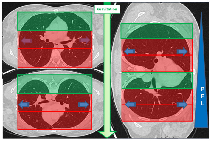
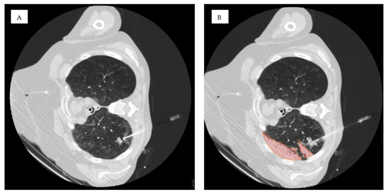
背景/目的:ct引导下肺活检中出现磨玻璃混浊(GGO)或在高度灌注、依赖的肺区域进行活检时出血的风险尚未系统研究。虽然已经研究了患者体位预防气胸的方法,但其在减少出血风险方面的作用仍未被探索。本研究旨在确定在ct引导下肺活检时,通路和依赖肺区活检中的GGOs是否是肺出血的危险因素。方法:回顾性分析单中心(2020-2023年)ct引导下的115例肺活检。患者根据介入后出血超过2cm(2级或更高)进行分类。我们使用卡方检验、Fisher精确检验和Mann-Whitney U检验评估了ggo在通路和活检位置(依赖与非依赖区域)的存在。采用单因素和多因素logistic回归分析评价肺出血的危险因素。结果:115例患者中有30例(26%)发生超过2cm的肺出血。在这些病例中,67%的患者在通路中发现了ggo (p < 0.01),在依赖肺区进行活检时出血发生率更高(63%对40%,p = 0.03)。多变量分析显示,通路内的ggo (OR 5.169, 95% CI 1.889-14.144, p = 0.001)和依赖区活检(OR 4.064, 95% CI 1.477-11.186, p < 0.001)分别增加出血风险。结论:ct引导下肺活检时,通路内的GGOs和依赖的肺区活检是出血的独立危险因素。
Ground-Glass Opacities in the Access Route and Biopsy in Highly Perfused Dependent Areas of the Lungs as Risk Factors for Pulmonary Hemorrhage During CT-Guided Lung Biopsy: A Retrospective Study.
Background/objectives: The risk of hemorrhage during CT-guided lung biopsy has not been systematically studied in cases where ground-glass opacities (GGO) are present in the access route or when biopsies are performed in highly perfused, dependent lung areas. While patient positioning has been studied for pneumothorax prevention, its role in minimizing hemorrhage risk remains unexplored. This study aimed to determine whether GGOs in the access route and biopsies in dependent lung areas are risk factors for pulmonary hemorrhage during CT-guided lung biopsy.
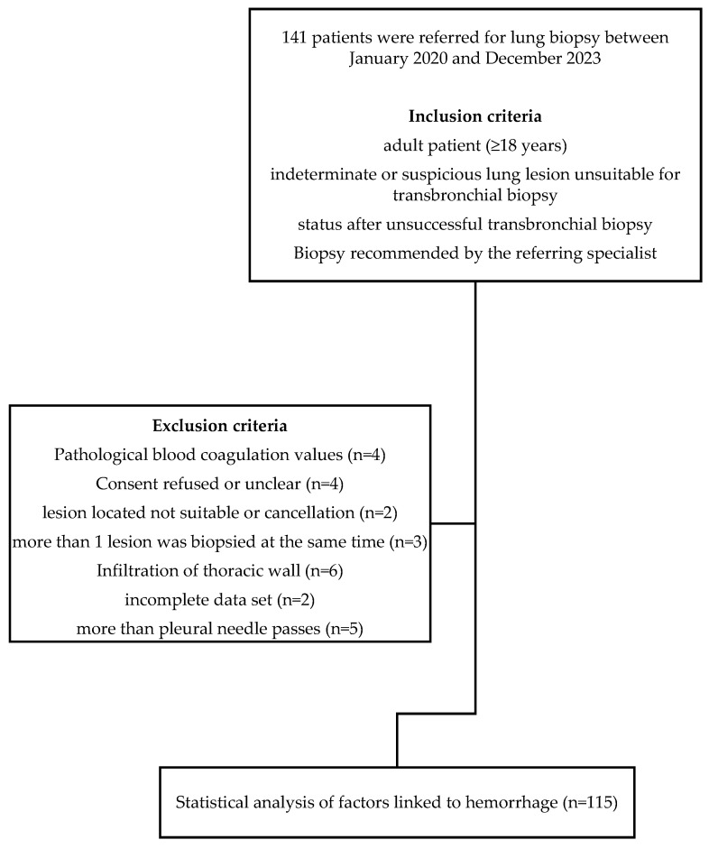
Methods: A retrospective analysis was conducted on 115 CT-guided lung biopsies performed at a single center (2020-2023). Patients were categorized based on post-interventional hemorrhage exceeding 2 cm (Grade 2 or higher). We evaluated the presence of GGOs in the access route and biopsy location (dependent vs. non-dependent areas) using chi square, Fisher's exact, and Mann-Whitney U tests. Univariate and multivariate logistic regression analyses were conducted to evaluate risk factors for pulmonary hemorrhage.
Results: Pulmonary hemorrhage beyond 2 cm occurred in 30 of 115 patients (26%). GGOs in the access route were identified in 67% of these cases (p < 0.01), and hemorrhage occurred more frequently when biopsies were performed in dependent lung areas (63% vs. 40%, p = 0.03). Multivariable analysis showed that GGOs in the access route (OR 5.169, 95% CI 1.889-14.144, p = 0.001) and biopsies in dependent areas (OR 4.064, 95% CI 1.477-11.186, p < 0.001) independently increased hemorrhage risk.
Conclusions: GGOs in the access route and dependent lung area biopsies are independent risk factors for hemorrhage during CT-guided lung biopsy.
TomographyMedicine-Radiology, Nuclear Medicine and Imaging
CiteScore
2.70
自引率
10.50%
发文量
222
期刊介绍:
TomographyTM publishes basic (technical and pre-clinical) and clinical scientific articles which involve the advancement of imaging technologies. Tomography encompasses studies that use single or multiple imaging modalities including for example CT, US, PET, SPECT, MR and hyperpolarization technologies, as well as optical modalities (i.e. bioluminescence, photoacoustic, endomicroscopy, fiber optic imaging and optical computed tomography) in basic sciences, engineering, preclinical and clinical medicine.
Tomography also welcomes studies involving exploration and refinement of contrast mechanisms and image-derived metrics within and across modalities toward the development of novel imaging probes for image-based feedback and intervention. The use of imaging in biology and medicine provides unparalleled opportunities to noninvasively interrogate tissues to obtain real-time dynamic and quantitative information required for diagnosis and response to interventions and to follow evolving pathological conditions. As multi-modal studies and the complexities of imaging technologies themselves are ever increasing to provide advanced information to scientists and clinicians.
Tomography provides a unique publication venue allowing investigators the opportunity to more precisely communicate integrated findings related to the diverse and heterogeneous features associated with underlying anatomical, physiological, functional, metabolic and molecular genetic activities of normal and diseased tissue. Thus Tomography publishes peer-reviewed articles which involve the broad use of imaging of any tissue and disease type including both preclinical and clinical investigations. In addition, hardware/software along with chemical and molecular probe advances are welcome as they are deemed to significantly contribute towards the long-term goal of improving the overall impact of imaging on scientific and clinical discovery.

 求助内容:
求助内容: 应助结果提醒方式:


