Maria Zaharieva Mutafchieva, Milena Nenkova Draganova, Georgi Tomchev Tomov
{"title":"口腔扁平苔藓的分子标记及其发病机制。","authors":"Maria Zaharieva Mutafchieva, Milena Nenkova Draganova, Georgi Tomchev Tomov","doi":"10.1007/s12105-025-01775-1","DOIUrl":null,"url":null,"abstract":"<p><strong>Purpose: </strong>Oral lichen planus (OLP) is a chronic inflammatory disease, characterized by immune-mediated basal keratinocyte apoptosis. In recent years the importance of programmed cell death for the tissue destruction in OLP has been disputed, while at the same time an increased proliferative index has been reported in the epithelium of these lesions. OLP is considered as a precancerous condition. This study investigated the expression of pro-apoptotic, anti-apoptotic and proliferative markers in OLP lesions in an attempt to understand more about the pathogenesis and malignant potential of the disease.</p><p><strong>Methods: </strong>Twenty patients with histologically confirmed OLP were compared to ten healthy controls through immunohistochemical analysis of the levels of p53, p63, bcl-2, Ki-67 and COX-2.</p><p><strong>Results: </strong>The results demonstrated significantly decreased expression of p63 in OLP lesions compared to normal oral mucosa. The levels of p53, bcl-2, Ki-67, and COX-2 were not significantly different from those in the control group. A significant association was found between p63 and Ki-67 (p = 0.001), as well as between p63 and p53 (p = 0.016). Expression of the inflammatory COX-2 and the apoptotic p53 appeared to be independent of each other (p = 0.44). The intensity of expression of any of the five analyzed markers was not related to the severity of the clinical manifestation.</p><p><strong>Conclusions: </strong>The obtained results suggest that apoptosis may not be the dominant mechanism in the disease's pathogenesis. Decreased expression of p63 on the other hand appears to play an important role. Among the possible effects of this protein deficiency are activation of programmed cell death, cell cycle arrest, cellular senescence, or anoikis; suppression of cell proliferation or changes in cell differentiation. The observed reduction in p63, Ki67 and bcl-2 levels predisposes to epithelial thinning, erosions and/or ulcers. For the presented OLP cohort, there was no molecular evidence of increased malignant potential of the lesions.</p>","PeriodicalId":47972,"journal":{"name":"Head & Neck Pathology","volume":"19 1","pages":"38"},"PeriodicalIF":4.1000,"publicationDate":"2025-03-26","publicationTypes":"Journal Article","fieldsOfStudy":null,"isOpenAccess":false,"openAccessPdf":"https://www.ncbi.nlm.nih.gov/pmc/articles/PMC11947335/pdf/","citationCount":"0","resultStr":"{\"title\":\"Molecular Markers in Oral Lichen Planus - Insight into Pathogenesis.\",\"authors\":\"Maria Zaharieva Mutafchieva, Milena Nenkova Draganova, Georgi Tomchev Tomov\",\"doi\":\"10.1007/s12105-025-01775-1\",\"DOIUrl\":null,\"url\":null,\"abstract\":\"<p><strong>Purpose: </strong>Oral lichen planus (OLP) is a chronic inflammatory disease, characterized by immune-mediated basal keratinocyte apoptosis. In recent years the importance of programmed cell death for the tissue destruction in OLP has been disputed, while at the same time an increased proliferative index has been reported in the epithelium of these lesions. OLP is considered as a precancerous condition. This study investigated the expression of pro-apoptotic, anti-apoptotic and proliferative markers in OLP lesions in an attempt to understand more about the pathogenesis and malignant potential of the disease.</p><p><strong>Methods: </strong>Twenty patients with histologically confirmed OLP were compared to ten healthy controls through immunohistochemical analysis of the levels of p53, p63, bcl-2, Ki-67 and COX-2.</p><p><strong>Results: </strong>The results demonstrated significantly decreased expression of p63 in OLP lesions compared to normal oral mucosa. The levels of p53, bcl-2, Ki-67, and COX-2 were not significantly different from those in the control group. A significant association was found between p63 and Ki-67 (p = 0.001), as well as between p63 and p53 (p = 0.016). Expression of the inflammatory COX-2 and the apoptotic p53 appeared to be independent of each other (p = 0.44). The intensity of expression of any of the five analyzed markers was not related to the severity of the clinical manifestation.</p><p><strong>Conclusions: </strong>The obtained results suggest that apoptosis may not be the dominant mechanism in the disease's pathogenesis. Decreased expression of p63 on the other hand appears to play an important role. Among the possible effects of this protein deficiency are activation of programmed cell death, cell cycle arrest, cellular senescence, or anoikis; suppression of cell proliferation or changes in cell differentiation. The observed reduction in p63, Ki67 and bcl-2 levels predisposes to epithelial thinning, erosions and/or ulcers. For the presented OLP cohort, there was no molecular evidence of increased malignant potential of the lesions.</p>\",\"PeriodicalId\":47972,\"journal\":{\"name\":\"Head & Neck Pathology\",\"volume\":\"19 1\",\"pages\":\"38\"},\"PeriodicalIF\":4.1000,\"publicationDate\":\"2025-03-26\",\"publicationTypes\":\"Journal Article\",\"fieldsOfStudy\":null,\"isOpenAccess\":false,\"openAccessPdf\":\"https://www.ncbi.nlm.nih.gov/pmc/articles/PMC11947335/pdf/\",\"citationCount\":\"0\",\"resultStr\":null,\"platform\":\"Semanticscholar\",\"paperid\":null,\"PeriodicalName\":\"Head & Neck Pathology\",\"FirstCategoryId\":\"1085\",\"ListUrlMain\":\"https://doi.org/10.1007/s12105-025-01775-1\",\"RegionNum\":0,\"RegionCategory\":null,\"ArticlePicture\":[],\"TitleCN\":null,\"AbstractTextCN\":null,\"PMCID\":null,\"EPubDate\":\"\",\"PubModel\":\"\",\"JCR\":\"Q2\",\"JCRName\":\"PATHOLOGY\",\"Score\":null,\"Total\":0}","platform":"Semanticscholar","paperid":null,"PeriodicalName":"Head & Neck Pathology","FirstCategoryId":"1085","ListUrlMain":"https://doi.org/10.1007/s12105-025-01775-1","RegionNum":0,"RegionCategory":null,"ArticlePicture":[],"TitleCN":null,"AbstractTextCN":null,"PMCID":null,"EPubDate":"","PubModel":"","JCR":"Q2","JCRName":"PATHOLOGY","Score":null,"Total":0}
Molecular Markers in Oral Lichen Planus - Insight into Pathogenesis.
Purpose: Oral lichen planus (OLP) is a chronic inflammatory disease, characterized by immune-mediated basal keratinocyte apoptosis. In recent years the importance of programmed cell death for the tissue destruction in OLP has been disputed, while at the same time an increased proliferative index has been reported in the epithelium of these lesions. OLP is considered as a precancerous condition. This study investigated the expression of pro-apoptotic, anti-apoptotic and proliferative markers in OLP lesions in an attempt to understand more about the pathogenesis and malignant potential of the disease.
Methods: Twenty patients with histologically confirmed OLP were compared to ten healthy controls through immunohistochemical analysis of the levels of p53, p63, bcl-2, Ki-67 and COX-2.
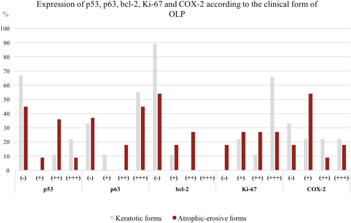
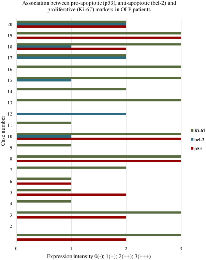
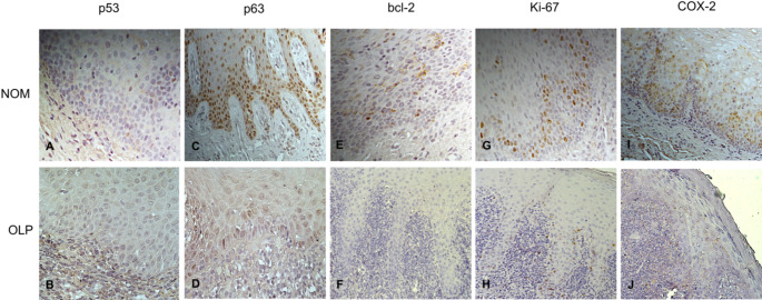
Results: The results demonstrated significantly decreased expression of p63 in OLP lesions compared to normal oral mucosa. The levels of p53, bcl-2, Ki-67, and COX-2 were not significantly different from those in the control group. A significant association was found between p63 and Ki-67 (p = 0.001), as well as between p63 and p53 (p = 0.016). Expression of the inflammatory COX-2 and the apoptotic p53 appeared to be independent of each other (p = 0.44). The intensity of expression of any of the five analyzed markers was not related to the severity of the clinical manifestation.
Conclusions: The obtained results suggest that apoptosis may not be the dominant mechanism in the disease's pathogenesis. Decreased expression of p63 on the other hand appears to play an important role. Among the possible effects of this protein deficiency are activation of programmed cell death, cell cycle arrest, cellular senescence, or anoikis; suppression of cell proliferation or changes in cell differentiation. The observed reduction in p63, Ki67 and bcl-2 levels predisposes to epithelial thinning, erosions and/or ulcers. For the presented OLP cohort, there was no molecular evidence of increased malignant potential of the lesions.
期刊介绍:
Head & Neck Pathology presents scholarly papers, reviews and symposia that cover the spectrum of human surgical pathology within the anatomic zones of the oral cavity, sinonasal tract, larynx, hypopharynx, salivary gland, ear and temporal bone, and neck.
The journal publishes rapid developments in new diagnostic criteria, intraoperative consultation, immunohistochemical studies, molecular techniques, genetic analyses, diagnostic aids, experimental pathology, cytology, radiographic imaging, and application of uniform terminology to allow practitioners to continue to maintain and expand their knowledge in the subspecialty of head and neck pathology. Coverage of practical application to daily clinical practice is supported with proceedings and symposia from international societies and academies devoted to this field.
Single-blind peer review
The journal follows a single-blind review procedure, where the reviewers are aware of the names and affiliations of the authors, but the reviewer reports provided to authors are anonymous. Single-blind peer review is the traditional model of peer review that many reviewers are comfortable with, and it facilitates a dispassionate critique of a manuscript.

 求助内容:
求助内容: 应助结果提醒方式:


