{"title":"基于多尺度图像和多特征融合框架的支气管超声图像诊断肺癌。","authors":"Huitao Wang, Takahiro Nakajima, Kohei Shikano, Yukihiro Nomura, Toshiya Nakaguchi","doi":"10.3390/tomography11030024","DOIUrl":null,"url":null,"abstract":"<p><p>Lung cancer is the leading cause of cancer-related deaths globally and ranks among the most common cancer types. Given its low overall five-year survival rate, early diagnosis and timely treatment are essential to improving patient outcomes. In recent years, advances in computer technology have enabled artificial intelligence to make groundbreaking progress in imaging-based lung cancer diagnosis. The primary aim of this study is to develop a computer-aided diagnosis (CAD) system for lung cancer using endobronchial ultrasonography (EBUS) images and deep learning algorithms to facilitate early detection and improve patient survival rates. We propose M3-Net, which is a multi-branch framework that integrates multiple features through an attention-based mechanism, enhancing diagnostic performance by providing more comprehensive information for lung cancer assessment. The framework was validated on a dataset of 95 patient cases, including 13 benign and 82 malignant cases. The dataset comprises 1140 EBUS images, with 540 images used for training, and 300 images each for the validation and test sets. The evaluation yielded the following results: accuracy of 0.76, F1-score of 0.75, AUC of 0.83, PPV of 0.80, NPV of 0.75, sensitivity of 0.72, and specificity of 0.80. These findings indicate that the proposed attention-based multi-feature fusion framework holds significant potential in assisting with lung cancer diagnosis.</p>","PeriodicalId":51330,"journal":{"name":"Tomography","volume":"11 3","pages":""},"PeriodicalIF":2.2000,"publicationDate":"2025-02-27","publicationTypes":"Journal Article","fieldsOfStudy":null,"isOpenAccess":false,"openAccessPdf":"https://www.ncbi.nlm.nih.gov/pmc/articles/PMC11945964/pdf/","citationCount":"0","resultStr":"{\"title\":\"Diagnosis of Lung Cancer Using Endobronchial Ultrasonography Image Based on Multi-Scale Image and Multi-Feature Fusion Framework.\",\"authors\":\"Huitao Wang, Takahiro Nakajima, Kohei Shikano, Yukihiro Nomura, Toshiya Nakaguchi\",\"doi\":\"10.3390/tomography11030024\",\"DOIUrl\":null,\"url\":null,\"abstract\":\"<p><p>Lung cancer is the leading cause of cancer-related deaths globally and ranks among the most common cancer types. Given its low overall five-year survival rate, early diagnosis and timely treatment are essential to improving patient outcomes. In recent years, advances in computer technology have enabled artificial intelligence to make groundbreaking progress in imaging-based lung cancer diagnosis. The primary aim of this study is to develop a computer-aided diagnosis (CAD) system for lung cancer using endobronchial ultrasonography (EBUS) images and deep learning algorithms to facilitate early detection and improve patient survival rates. We propose M3-Net, which is a multi-branch framework that integrates multiple features through an attention-based mechanism, enhancing diagnostic performance by providing more comprehensive information for lung cancer assessment. The framework was validated on a dataset of 95 patient cases, including 13 benign and 82 malignant cases. The dataset comprises 1140 EBUS images, with 540 images used for training, and 300 images each for the validation and test sets. The evaluation yielded the following results: accuracy of 0.76, F1-score of 0.75, AUC of 0.83, PPV of 0.80, NPV of 0.75, sensitivity of 0.72, and specificity of 0.80. These findings indicate that the proposed attention-based multi-feature fusion framework holds significant potential in assisting with lung cancer diagnosis.</p>\",\"PeriodicalId\":51330,\"journal\":{\"name\":\"Tomography\",\"volume\":\"11 3\",\"pages\":\"\"},\"PeriodicalIF\":2.2000,\"publicationDate\":\"2025-02-27\",\"publicationTypes\":\"Journal Article\",\"fieldsOfStudy\":null,\"isOpenAccess\":false,\"openAccessPdf\":\"https://www.ncbi.nlm.nih.gov/pmc/articles/PMC11945964/pdf/\",\"citationCount\":\"0\",\"resultStr\":null,\"platform\":\"Semanticscholar\",\"paperid\":null,\"PeriodicalName\":\"Tomography\",\"FirstCategoryId\":\"3\",\"ListUrlMain\":\"https://doi.org/10.3390/tomography11030024\",\"RegionNum\":4,\"RegionCategory\":\"医学\",\"ArticlePicture\":[],\"TitleCN\":null,\"AbstractTextCN\":null,\"PMCID\":null,\"EPubDate\":\"\",\"PubModel\":\"\",\"JCR\":\"Q2\",\"JCRName\":\"RADIOLOGY, NUCLEAR MEDICINE & MEDICAL IMAGING\",\"Score\":null,\"Total\":0}","platform":"Semanticscholar","paperid":null,"PeriodicalName":"Tomography","FirstCategoryId":"3","ListUrlMain":"https://doi.org/10.3390/tomography11030024","RegionNum":4,"RegionCategory":"医学","ArticlePicture":[],"TitleCN":null,"AbstractTextCN":null,"PMCID":null,"EPubDate":"","PubModel":"","JCR":"Q2","JCRName":"RADIOLOGY, NUCLEAR MEDICINE & MEDICAL IMAGING","Score":null,"Total":0}
Diagnosis of Lung Cancer Using Endobronchial Ultrasonography Image Based on Multi-Scale Image and Multi-Feature Fusion Framework.
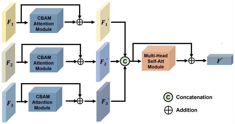
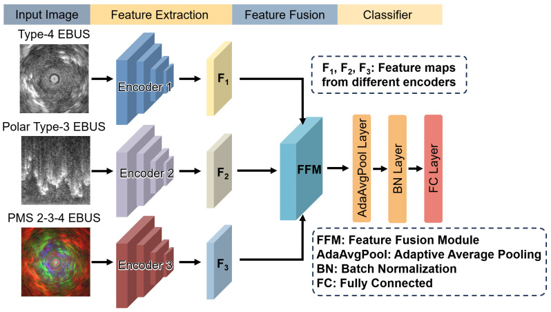
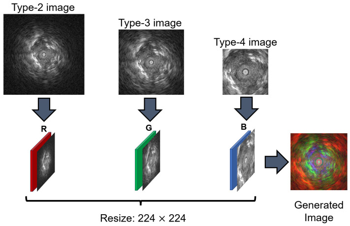
Lung cancer is the leading cause of cancer-related deaths globally and ranks among the most common cancer types. Given its low overall five-year survival rate, early diagnosis and timely treatment are essential to improving patient outcomes. In recent years, advances in computer technology have enabled artificial intelligence to make groundbreaking progress in imaging-based lung cancer diagnosis. The primary aim of this study is to develop a computer-aided diagnosis (CAD) system for lung cancer using endobronchial ultrasonography (EBUS) images and deep learning algorithms to facilitate early detection and improve patient survival rates. We propose M3-Net, which is a multi-branch framework that integrates multiple features through an attention-based mechanism, enhancing diagnostic performance by providing more comprehensive information for lung cancer assessment. The framework was validated on a dataset of 95 patient cases, including 13 benign and 82 malignant cases. The dataset comprises 1140 EBUS images, with 540 images used for training, and 300 images each for the validation and test sets. The evaluation yielded the following results: accuracy of 0.76, F1-score of 0.75, AUC of 0.83, PPV of 0.80, NPV of 0.75, sensitivity of 0.72, and specificity of 0.80. These findings indicate that the proposed attention-based multi-feature fusion framework holds significant potential in assisting with lung cancer diagnosis.
TomographyMedicine-Radiology, Nuclear Medicine and Imaging
CiteScore
2.70
自引率
10.50%
发文量
222
期刊介绍:
TomographyTM publishes basic (technical and pre-clinical) and clinical scientific articles which involve the advancement of imaging technologies. Tomography encompasses studies that use single or multiple imaging modalities including for example CT, US, PET, SPECT, MR and hyperpolarization technologies, as well as optical modalities (i.e. bioluminescence, photoacoustic, endomicroscopy, fiber optic imaging and optical computed tomography) in basic sciences, engineering, preclinical and clinical medicine.
Tomography also welcomes studies involving exploration and refinement of contrast mechanisms and image-derived metrics within and across modalities toward the development of novel imaging probes for image-based feedback and intervention. The use of imaging in biology and medicine provides unparalleled opportunities to noninvasively interrogate tissues to obtain real-time dynamic and quantitative information required for diagnosis and response to interventions and to follow evolving pathological conditions. As multi-modal studies and the complexities of imaging technologies themselves are ever increasing to provide advanced information to scientists and clinicians.
Tomography provides a unique publication venue allowing investigators the opportunity to more precisely communicate integrated findings related to the diverse and heterogeneous features associated with underlying anatomical, physiological, functional, metabolic and molecular genetic activities of normal and diseased tissue. Thus Tomography publishes peer-reviewed articles which involve the broad use of imaging of any tissue and disease type including both preclinical and clinical investigations. In addition, hardware/software along with chemical and molecular probe advances are welcome as they are deemed to significantly contribute towards the long-term goal of improving the overall impact of imaging on scientific and clinical discovery.

 求助内容:
求助内容: 应助结果提醒方式:


