{"title":"基于深度学习的图像质量增强方法对数字bgo PET/CT系统18F-FDG全身检查的影响","authors":"Kenta Miwa, Shin Yamagishi, Shun Kamitaki, Kouichi Anraku, Shun Sato, Tensho Yamao, Noriaki Miyaji, Kaito Wachi, Naochika Akiya, Kei Wagatsuma, Kazuhiro Oguchi","doi":"10.1186/s40658-025-00742-7","DOIUrl":null,"url":null,"abstract":"<p><strong>Background: </strong>The digital-BGO PET/CT system, Omni Legend 32, incorporates modified block sequential regularized expectation maximization (BSREM) image reconstruction and a deep learning-based time-of-flight (TOF)-like image quality enhancement process called Precision DL (PDL). The present study aimed to define the fundamental characteristics of PDL using phantom and clinical images.</p><p><strong>Methods: </strong>A NEMA IEC body phantom was scanned using the Omni Legend 32 PET/CT system. All PET/CT images were acquired over 60 and 90 s per bed position, with a 384 × 384 matrix. Phantom images were reconstructed using OSEM + PSF and BSREM at β values of 100-1,000, combined with low (LPDL), medium (MPDL), and high (HPDL) PDL. We evaluated contrast recovery, background variability, and the contrast-to-noise ratio (CNR) of a 10 mm hot sphere. Thirty clinical whole-body <sup>18</sup>F-FDG PET/CT examinations were included. Clinical images were reconstructed using OSEM + PSF and BSREM at β values of 200, 300, 400, 500, and 600, determined based on findings from the phantom study, combined with the three PDL models. Noise levels, mean SUV (SUV<sub>mean</sub>), and the signal-to-noise ratio (SNR) of the liver as well as signal-to-background ratios (SBR) and maximum SUV (SUV<sub>max</sub>) of lesions were evaluated. Two blinded readers evaluated visual image quality and rated several aspects to complement the analysis.</p><p><strong>Results: </strong>Contrast recovery and background variability decreased as the β value increased. This trend was consistent even when PDL processing was added to BSREM. Increased strength of the PDL models led to higher CNR. Noise levels decreased as a function of increasing β values in BSREM, resulting in a higher SNR, but lower SBR. Combining PDL with BSREM resulted in all β values producing better results in terms of noise, SBR, and SNR than OSEM + PSF. As the PDL increased (LPDL < MPDL < HPDL), noise levels, SBR, and SNR became higher. The β values of 400, 200, 300, and 300 for BSREM, LPDL, MPDL, and HPDL, respectively, resulted in noise equivalent to OSEM + PSF but significantly increased the SUV<sub>max</sub> (9%, 15%, 18%, and 27%), SBR (16%, 17%, 20%, and 32%), and SNR (17%, 19%, 31%, and 36%), respectively. The visual evaluation of image quality yielded similar scores across BSREM + PDL reconstructions, although BSREM with β = 600 combined with MPDL delivered the best overall image quality and total mean score.</p><p><strong>Conclusion: </strong>The combination of BSREM and PDL significantly enhanced the SUV<sub>max</sub> of lesions and image quality compared with OSEM + PSF. A combination of BSREM at β values of 500-600 and MPDL is recommended for oncological whole-body PET/CT imaging when using PDL on the Omni Legend.</p>","PeriodicalId":11559,"journal":{"name":"EJNMMI Physics","volume":"12 1","pages":"29"},"PeriodicalIF":3.2000,"publicationDate":"2025-03-28","publicationTypes":"Journal Article","fieldsOfStudy":null,"isOpenAccess":false,"openAccessPdf":"https://www.ncbi.nlm.nih.gov/pmc/articles/PMC11950486/pdf/","citationCount":"0","resultStr":"{\"title\":\"Effects of a deep learning-based image quality enhancement method on a digital-BGO PET/CT system for <sup>18</sup>F-FDG whole-body examination.\",\"authors\":\"Kenta Miwa, Shin Yamagishi, Shun Kamitaki, Kouichi Anraku, Shun Sato, Tensho Yamao, Noriaki Miyaji, Kaito Wachi, Naochika Akiya, Kei Wagatsuma, Kazuhiro Oguchi\",\"doi\":\"10.1186/s40658-025-00742-7\",\"DOIUrl\":null,\"url\":null,\"abstract\":\"<p><strong>Background: </strong>The digital-BGO PET/CT system, Omni Legend 32, incorporates modified block sequential regularized expectation maximization (BSREM) image reconstruction and a deep learning-based time-of-flight (TOF)-like image quality enhancement process called Precision DL (PDL). The present study aimed to define the fundamental characteristics of PDL using phantom and clinical images.</p><p><strong>Methods: </strong>A NEMA IEC body phantom was scanned using the Omni Legend 32 PET/CT system. All PET/CT images were acquired over 60 and 90 s per bed position, with a 384 × 384 matrix. Phantom images were reconstructed using OSEM + PSF and BSREM at β values of 100-1,000, combined with low (LPDL), medium (MPDL), and high (HPDL) PDL. We evaluated contrast recovery, background variability, and the contrast-to-noise ratio (CNR) of a 10 mm hot sphere. Thirty clinical whole-body <sup>18</sup>F-FDG PET/CT examinations were included. Clinical images were reconstructed using OSEM + PSF and BSREM at β values of 200, 300, 400, 500, and 600, determined based on findings from the phantom study, combined with the three PDL models. Noise levels, mean SUV (SUV<sub>mean</sub>), and the signal-to-noise ratio (SNR) of the liver as well as signal-to-background ratios (SBR) and maximum SUV (SUV<sub>max</sub>) of lesions were evaluated. Two blinded readers evaluated visual image quality and rated several aspects to complement the analysis.</p><p><strong>Results: </strong>Contrast recovery and background variability decreased as the β value increased. This trend was consistent even when PDL processing was added to BSREM. Increased strength of the PDL models led to higher CNR. Noise levels decreased as a function of increasing β values in BSREM, resulting in a higher SNR, but lower SBR. Combining PDL with BSREM resulted in all β values producing better results in terms of noise, SBR, and SNR than OSEM + PSF. As the PDL increased (LPDL < MPDL < HPDL), noise levels, SBR, and SNR became higher. The β values of 400, 200, 300, and 300 for BSREM, LPDL, MPDL, and HPDL, respectively, resulted in noise equivalent to OSEM + PSF but significantly increased the SUV<sub>max</sub> (9%, 15%, 18%, and 27%), SBR (16%, 17%, 20%, and 32%), and SNR (17%, 19%, 31%, and 36%), respectively. The visual evaluation of image quality yielded similar scores across BSREM + PDL reconstructions, although BSREM with β = 600 combined with MPDL delivered the best overall image quality and total mean score.</p><p><strong>Conclusion: </strong>The combination of BSREM and PDL significantly enhanced the SUV<sub>max</sub> of lesions and image quality compared with OSEM + PSF. A combination of BSREM at β values of 500-600 and MPDL is recommended for oncological whole-body PET/CT imaging when using PDL on the Omni Legend.</p>\",\"PeriodicalId\":11559,\"journal\":{\"name\":\"EJNMMI Physics\",\"volume\":\"12 1\",\"pages\":\"29\"},\"PeriodicalIF\":3.2000,\"publicationDate\":\"2025-03-28\",\"publicationTypes\":\"Journal Article\",\"fieldsOfStudy\":null,\"isOpenAccess\":false,\"openAccessPdf\":\"https://www.ncbi.nlm.nih.gov/pmc/articles/PMC11950486/pdf/\",\"citationCount\":\"0\",\"resultStr\":null,\"platform\":\"Semanticscholar\",\"paperid\":null,\"PeriodicalName\":\"EJNMMI Physics\",\"FirstCategoryId\":\"3\",\"ListUrlMain\":\"https://doi.org/10.1186/s40658-025-00742-7\",\"RegionNum\":2,\"RegionCategory\":\"医学\",\"ArticlePicture\":[],\"TitleCN\":null,\"AbstractTextCN\":null,\"PMCID\":null,\"EPubDate\":\"\",\"PubModel\":\"\",\"JCR\":\"Q2\",\"JCRName\":\"RADIOLOGY, NUCLEAR MEDICINE & MEDICAL IMAGING\",\"Score\":null,\"Total\":0}","platform":"Semanticscholar","paperid":null,"PeriodicalName":"EJNMMI Physics","FirstCategoryId":"3","ListUrlMain":"https://doi.org/10.1186/s40658-025-00742-7","RegionNum":2,"RegionCategory":"医学","ArticlePicture":[],"TitleCN":null,"AbstractTextCN":null,"PMCID":null,"EPubDate":"","PubModel":"","JCR":"Q2","JCRName":"RADIOLOGY, NUCLEAR MEDICINE & MEDICAL IMAGING","Score":null,"Total":0}
Effects of a deep learning-based image quality enhancement method on a digital-BGO PET/CT system for 18F-FDG whole-body examination.
Background: The digital-BGO PET/CT system, Omni Legend 32, incorporates modified block sequential regularized expectation maximization (BSREM) image reconstruction and a deep learning-based time-of-flight (TOF)-like image quality enhancement process called Precision DL (PDL). The present study aimed to define the fundamental characteristics of PDL using phantom and clinical images.
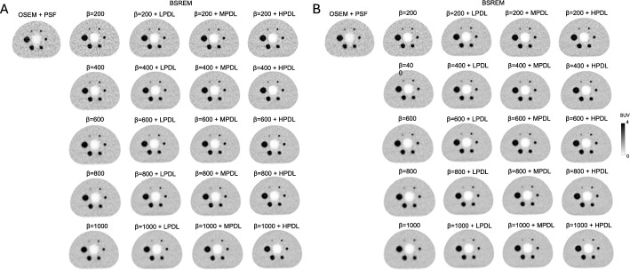
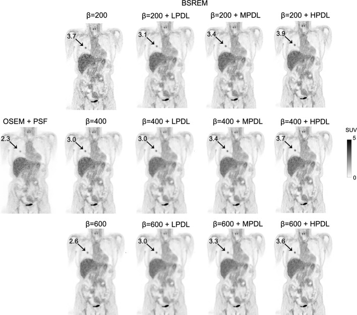
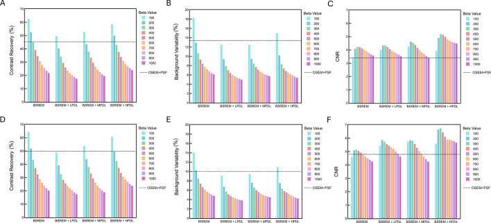
Methods: A NEMA IEC body phantom was scanned using the Omni Legend 32 PET/CT system. All PET/CT images were acquired over 60 and 90 s per bed position, with a 384 × 384 matrix. Phantom images were reconstructed using OSEM + PSF and BSREM at β values of 100-1,000, combined with low (LPDL), medium (MPDL), and high (HPDL) PDL. We evaluated contrast recovery, background variability, and the contrast-to-noise ratio (CNR) of a 10 mm hot sphere. Thirty clinical whole-body 18F-FDG PET/CT examinations were included. Clinical images were reconstructed using OSEM + PSF and BSREM at β values of 200, 300, 400, 500, and 600, determined based on findings from the phantom study, combined with the three PDL models. Noise levels, mean SUV (SUVmean), and the signal-to-noise ratio (SNR) of the liver as well as signal-to-background ratios (SBR) and maximum SUV (SUVmax) of lesions were evaluated. Two blinded readers evaluated visual image quality and rated several aspects to complement the analysis.
Results: Contrast recovery and background variability decreased as the β value increased. This trend was consistent even when PDL processing was added to BSREM. Increased strength of the PDL models led to higher CNR. Noise levels decreased as a function of increasing β values in BSREM, resulting in a higher SNR, but lower SBR. Combining PDL with BSREM resulted in all β values producing better results in terms of noise, SBR, and SNR than OSEM + PSF. As the PDL increased (LPDL < MPDL < HPDL), noise levels, SBR, and SNR became higher. The β values of 400, 200, 300, and 300 for BSREM, LPDL, MPDL, and HPDL, respectively, resulted in noise equivalent to OSEM + PSF but significantly increased the SUVmax (9%, 15%, 18%, and 27%), SBR (16%, 17%, 20%, and 32%), and SNR (17%, 19%, 31%, and 36%), respectively. The visual evaluation of image quality yielded similar scores across BSREM + PDL reconstructions, although BSREM with β = 600 combined with MPDL delivered the best overall image quality and total mean score.
Conclusion: The combination of BSREM and PDL significantly enhanced the SUVmax of lesions and image quality compared with OSEM + PSF. A combination of BSREM at β values of 500-600 and MPDL is recommended for oncological whole-body PET/CT imaging when using PDL on the Omni Legend.
期刊介绍:
EJNMMI Physics is an international platform for scientists, users and adopters of nuclear medicine with a particular interest in physics matters. As a companion journal to the European Journal of Nuclear Medicine and Molecular Imaging, this journal has a multi-disciplinary approach and welcomes original materials and studies with a focus on applied physics and mathematics as well as imaging systems engineering and prototyping in nuclear medicine. This includes physics-driven approaches or algorithms supported by physics that foster early clinical adoption of nuclear medicine imaging and therapy.

 求助内容:
求助内容: 应助结果提醒方式:


