{"title":"病例报告:骨扫描在检测肋病中的作用。","authors":"Abel Dambrain, Clément Bouron, Franck Lacoeuille","doi":"10.3389/fnume.2025.1527159","DOIUrl":null,"url":null,"abstract":"<p><p>In this case, we report the usefulness of bone scintigraphy in evaluating osteoarticular pain when the diagnosis is unclear after standard morphological imaging. A 24-year-old male patient exhibited mild left tibial pain that had been intensifying over a period of 2 years. The initial radiological evaluation suggested a diagnosis of pediatric tibial bone marrow osteosclerosis associated with periostitis, based on standard radiographs and MRI. However, a complementary bone scan was required for confirmation and showed moderate hyperemia and severe hyperfixation of the tibial lesion along with similar lesions on the left femur, both humeri, and the right ulna. These new findings led to a diagnosis of Ribbing disease, a rare sclerosing bone dysplasia.</p>","PeriodicalId":73095,"journal":{"name":"Frontiers in nuclear medicine (Lausanne, Switzerland)","volume":"5 ","pages":"1527159"},"PeriodicalIF":1.4000,"publicationDate":"2025-03-12","publicationTypes":"Journal Article","fieldsOfStudy":null,"isOpenAccess":false,"openAccessPdf":"https://www.ncbi.nlm.nih.gov/pmc/articles/PMC11936802/pdf/","citationCount":"0","resultStr":"{\"title\":\"Case Report: The role of bone scans in detecting Ribbing disease.\",\"authors\":\"Abel Dambrain, Clément Bouron, Franck Lacoeuille\",\"doi\":\"10.3389/fnume.2025.1527159\",\"DOIUrl\":null,\"url\":null,\"abstract\":\"<p><p>In this case, we report the usefulness of bone scintigraphy in evaluating osteoarticular pain when the diagnosis is unclear after standard morphological imaging. A 24-year-old male patient exhibited mild left tibial pain that had been intensifying over a period of 2 years. The initial radiological evaluation suggested a diagnosis of pediatric tibial bone marrow osteosclerosis associated with periostitis, based on standard radiographs and MRI. However, a complementary bone scan was required for confirmation and showed moderate hyperemia and severe hyperfixation of the tibial lesion along with similar lesions on the left femur, both humeri, and the right ulna. These new findings led to a diagnosis of Ribbing disease, a rare sclerosing bone dysplasia.</p>\",\"PeriodicalId\":73095,\"journal\":{\"name\":\"Frontiers in nuclear medicine (Lausanne, Switzerland)\",\"volume\":\"5 \",\"pages\":\"1527159\"},\"PeriodicalIF\":1.4000,\"publicationDate\":\"2025-03-12\",\"publicationTypes\":\"Journal Article\",\"fieldsOfStudy\":null,\"isOpenAccess\":false,\"openAccessPdf\":\"https://www.ncbi.nlm.nih.gov/pmc/articles/PMC11936802/pdf/\",\"citationCount\":\"0\",\"resultStr\":null,\"platform\":\"Semanticscholar\",\"paperid\":null,\"PeriodicalName\":\"Frontiers in nuclear medicine (Lausanne, Switzerland)\",\"FirstCategoryId\":\"1085\",\"ListUrlMain\":\"https://doi.org/10.3389/fnume.2025.1527159\",\"RegionNum\":0,\"RegionCategory\":null,\"ArticlePicture\":[],\"TitleCN\":null,\"AbstractTextCN\":null,\"PMCID\":null,\"EPubDate\":\"2025/1/1 0:00:00\",\"PubModel\":\"eCollection\",\"JCR\":\"\",\"JCRName\":\"\",\"Score\":null,\"Total\":0}","platform":"Semanticscholar","paperid":null,"PeriodicalName":"Frontiers in nuclear medicine (Lausanne, Switzerland)","FirstCategoryId":"1085","ListUrlMain":"https://doi.org/10.3389/fnume.2025.1527159","RegionNum":0,"RegionCategory":null,"ArticlePicture":[],"TitleCN":null,"AbstractTextCN":null,"PMCID":null,"EPubDate":"2025/1/1 0:00:00","PubModel":"eCollection","JCR":"","JCRName":"","Score":null,"Total":0}
Case Report: The role of bone scans in detecting Ribbing disease.
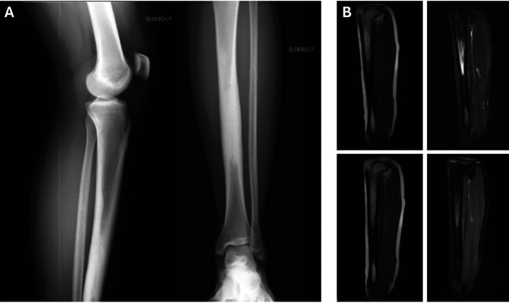
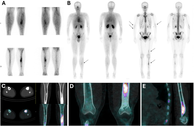
In this case, we report the usefulness of bone scintigraphy in evaluating osteoarticular pain when the diagnosis is unclear after standard morphological imaging. A 24-year-old male patient exhibited mild left tibial pain that had been intensifying over a period of 2 years. The initial radiological evaluation suggested a diagnosis of pediatric tibial bone marrow osteosclerosis associated with periostitis, based on standard radiographs and MRI. However, a complementary bone scan was required for confirmation and showed moderate hyperemia and severe hyperfixation of the tibial lesion along with similar lesions on the left femur, both humeri, and the right ulna. These new findings led to a diagnosis of Ribbing disease, a rare sclerosing bone dysplasia.

 求助内容:
求助内容: 应助结果提醒方式:


