{"title":"肉桂醛通过TAK1-p38MAPK-NLRP3通路对室性心律失常的影响及分子机制","authors":"Guoping Ma, Mian Li, Wanyue Yang, Hai Wang, Xue Tian, Yajuan Yin, Yida Tang, Wenjie Liang","doi":"10.1007/s00380-025-02529-3","DOIUrl":null,"url":null,"abstract":"<p><p>Based on the transforming growth factor β-activated kinase 1 (TAK1)-p38 mitogen-activated protein kinase (p38MAPK)-nucleotide-binding oligo-like receptor protein 3 (NLRP3) signalling pathway, the protective effect and mechanism of isoproterennaline (ISO)-induced cinnamaldehyde on inflammatory injury in ventricular rats were investigated. Fifty male SPF SD rats were randomly assigned to the normal group, model group, propranolol group, cinnamaldehyde low-dose group or cinnamaldehyde high-dose group. The ventricular arrhythmia model was constructed using the \"6 + 1\" ISO injection method. The rats in the propranolol group were given propranolol 15 mg·(kg d)<sup>-1</sup>, those in the low and high-dose groups were given cinnamaldehyde 20 mg·(kg d)<sup>-1</sup> and 50 mg·(kg d)<sup>-1</sup>, respectively, and those in the control and model groups received an equal volume of 0.9% NaCl solution. Changes in the serum troponin (cTnI), creatine kinase isoenzyme (CK-MB), and interleukin-1β (IL-1β) levels in SD rats were determined by ELISA. HE staining was used to observe the tissue morphology of heart disease. The mRNA expression of IL-1β and NLRP3 was determined by RT‒PCR. Mitochondrial damage was observed by transmission electron microscopy. The expression of reactive oxygen species (ROS) was detected by immunofluorescence. Western blot or immunohistochemical detection of the protein expression of IL-1β, NLRP3, TAK1, phospho-TAK1 (p-TAK1), p38MAPK, phospho-p38MAPK (p-p38MAPK), nuclear factor-κB (NF-κB),and phospho-NF-κB (p-NF-κB) was also performed. Data analysis was performed using SPSS 25.0 software. In the control SD rats, there were no obvious ventricular arrhythmias on ECG, the cardiac tissue and mitochondria were basically normal, the serum IL-1β level was low, and the expression of myocardial IL-1β, NLRP3, ROS, p-TAK1, p-p38MAPK and p-NF-κB was weak. Compared with the control group, the model group of SD rats had significant increases in ventricular arrhythmia and arrhythmia scores according to ECG (P < 0.01). Myocardial histopathological injury, cardiac weight index (HWI) and increases in serum cTnI and CK-MB levels were detected (P < 0.01). Additionally, mitochondrial damage in myocardial tissue, increased ROS fluorescence intensity, and elevated expression of myocardial p-TAK1, p-p38MAPK and p-NF-κB were detected(P < 0.01). The protein and mRNA expression of inflammation-related factors NLRP3 and IL-1β were increased (P < 0.01 or P < 0.05). Compared with those in the model group, the arrhythmia scores were decreased in the three treatment groups (P < 0.01 or P < 0.05). Cardiac histopathological morphology was significantly improved, and HWI and myocardial injury-related indicators were decreased(P < 0.01 or P < 0.05). Damaged mitochondria were significantly improved, and the expression of ROS, p-TAK1, p-p38MAPK, and p-NF-κB were decreased. The expression of inflammation-related factors in serum and myocardial tissue was decreased (P < 0.01 or P < 0.05). TAK1-p38MAPK-NLRP3 signalling is enhanced in SD rats with ventricular arrhythmia. Cinnamaldehyde can regulate TAK1-p38MAPK-NLRP3 signalling, reduce cardiomyocyte pyroptosis, antagonize myocardial inflammatory injury and protect cardiomyocytes by inhibiting oxidative stress.</p>","PeriodicalId":12940,"journal":{"name":"Heart and Vessels","volume":" ","pages":"846-861"},"PeriodicalIF":1.5000,"publicationDate":"2025-09-01","publicationTypes":"Journal Article","fieldsOfStudy":null,"isOpenAccess":false,"openAccessPdf":"https://www.ncbi.nlm.nih.gov/pmc/articles/PMC12380895/pdf/","citationCount":"0","resultStr":"{\"title\":\"Influence and molecular mechanism of cinnamaldehyde against ventricular arrhythmia via the TAK1-p38MAPK-NLRP3 pathway.\",\"authors\":\"Guoping Ma, Mian Li, Wanyue Yang, Hai Wang, Xue Tian, Yajuan Yin, Yida Tang, Wenjie Liang\",\"doi\":\"10.1007/s00380-025-02529-3\",\"DOIUrl\":null,\"url\":null,\"abstract\":\"<p><p>Based on the transforming growth factor β-activated kinase 1 (TAK1)-p38 mitogen-activated protein kinase (p38MAPK)-nucleotide-binding oligo-like receptor protein 3 (NLRP3) signalling pathway, the protective effect and mechanism of isoproterennaline (ISO)-induced cinnamaldehyde on inflammatory injury in ventricular rats were investigated. Fifty male SPF SD rats were randomly assigned to the normal group, model group, propranolol group, cinnamaldehyde low-dose group or cinnamaldehyde high-dose group. The ventricular arrhythmia model was constructed using the \\\"6 + 1\\\" ISO injection method. The rats in the propranolol group were given propranolol 15 mg·(kg d)<sup>-1</sup>, those in the low and high-dose groups were given cinnamaldehyde 20 mg·(kg d)<sup>-1</sup> and 50 mg·(kg d)<sup>-1</sup>, respectively, and those in the control and model groups received an equal volume of 0.9% NaCl solution. Changes in the serum troponin (cTnI), creatine kinase isoenzyme (CK-MB), and interleukin-1β (IL-1β) levels in SD rats were determined by ELISA. HE staining was used to observe the tissue morphology of heart disease. The mRNA expression of IL-1β and NLRP3 was determined by RT‒PCR. Mitochondrial damage was observed by transmission electron microscopy. The expression of reactive oxygen species (ROS) was detected by immunofluorescence. Western blot or immunohistochemical detection of the protein expression of IL-1β, NLRP3, TAK1, phospho-TAK1 (p-TAK1), p38MAPK, phospho-p38MAPK (p-p38MAPK), nuclear factor-κB (NF-κB),and phospho-NF-κB (p-NF-κB) was also performed. Data analysis was performed using SPSS 25.0 software. In the control SD rats, there were no obvious ventricular arrhythmias on ECG, the cardiac tissue and mitochondria were basically normal, the serum IL-1β level was low, and the expression of myocardial IL-1β, NLRP3, ROS, p-TAK1, p-p38MAPK and p-NF-κB was weak. Compared with the control group, the model group of SD rats had significant increases in ventricular arrhythmia and arrhythmia scores according to ECG (P < 0.01). Myocardial histopathological injury, cardiac weight index (HWI) and increases in serum cTnI and CK-MB levels were detected (P < 0.01). Additionally, mitochondrial damage in myocardial tissue, increased ROS fluorescence intensity, and elevated expression of myocardial p-TAK1, p-p38MAPK and p-NF-κB were detected(P < 0.01). The protein and mRNA expression of inflammation-related factors NLRP3 and IL-1β were increased (P < 0.01 or P < 0.05). Compared with those in the model group, the arrhythmia scores were decreased in the three treatment groups (P < 0.01 or P < 0.05). Cardiac histopathological morphology was significantly improved, and HWI and myocardial injury-related indicators were decreased(P < 0.01 or P < 0.05). Damaged mitochondria were significantly improved, and the expression of ROS, p-TAK1, p-p38MAPK, and p-NF-κB were decreased. The expression of inflammation-related factors in serum and myocardial tissue was decreased (P < 0.01 or P < 0.05). TAK1-p38MAPK-NLRP3 signalling is enhanced in SD rats with ventricular arrhythmia. Cinnamaldehyde can regulate TAK1-p38MAPK-NLRP3 signalling, reduce cardiomyocyte pyroptosis, antagonize myocardial inflammatory injury and protect cardiomyocytes by inhibiting oxidative stress.</p>\",\"PeriodicalId\":12940,\"journal\":{\"name\":\"Heart and Vessels\",\"volume\":\" \",\"pages\":\"846-861\"},\"PeriodicalIF\":1.5000,\"publicationDate\":\"2025-09-01\",\"publicationTypes\":\"Journal Article\",\"fieldsOfStudy\":null,\"isOpenAccess\":false,\"openAccessPdf\":\"https://www.ncbi.nlm.nih.gov/pmc/articles/PMC12380895/pdf/\",\"citationCount\":\"0\",\"resultStr\":null,\"platform\":\"Semanticscholar\",\"paperid\":null,\"PeriodicalName\":\"Heart and Vessels\",\"FirstCategoryId\":\"3\",\"ListUrlMain\":\"https://doi.org/10.1007/s00380-025-02529-3\",\"RegionNum\":4,\"RegionCategory\":\"医学\",\"ArticlePicture\":[],\"TitleCN\":null,\"AbstractTextCN\":null,\"PMCID\":null,\"EPubDate\":\"2025/3/27 0:00:00\",\"PubModel\":\"Epub\",\"JCR\":\"Q3\",\"JCRName\":\"CARDIAC & CARDIOVASCULAR SYSTEMS\",\"Score\":null,\"Total\":0}","platform":"Semanticscholar","paperid":null,"PeriodicalName":"Heart and Vessels","FirstCategoryId":"3","ListUrlMain":"https://doi.org/10.1007/s00380-025-02529-3","RegionNum":4,"RegionCategory":"医学","ArticlePicture":[],"TitleCN":null,"AbstractTextCN":null,"PMCID":null,"EPubDate":"2025/3/27 0:00:00","PubModel":"Epub","JCR":"Q3","JCRName":"CARDIAC & CARDIOVASCULAR SYSTEMS","Score":null,"Total":0}
Influence and molecular mechanism of cinnamaldehyde against ventricular arrhythmia via the TAK1-p38MAPK-NLRP3 pathway.
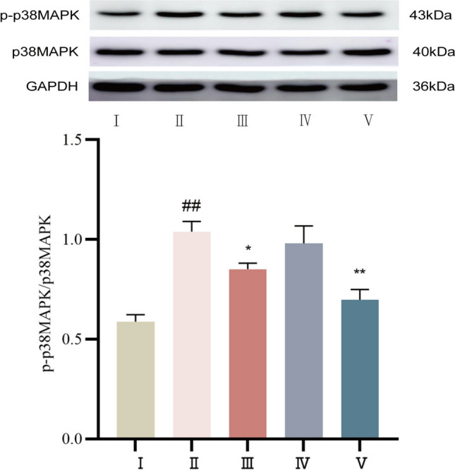
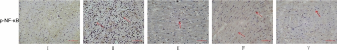
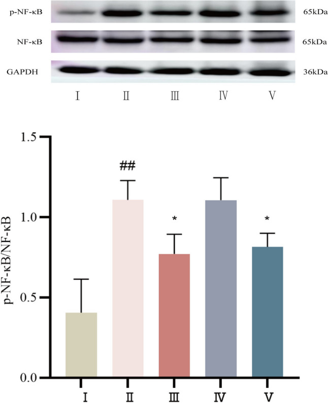
Based on the transforming growth factor β-activated kinase 1 (TAK1)-p38 mitogen-activated protein kinase (p38MAPK)-nucleotide-binding oligo-like receptor protein 3 (NLRP3) signalling pathway, the protective effect and mechanism of isoproterennaline (ISO)-induced cinnamaldehyde on inflammatory injury in ventricular rats were investigated. Fifty male SPF SD rats were randomly assigned to the normal group, model group, propranolol group, cinnamaldehyde low-dose group or cinnamaldehyde high-dose group. The ventricular arrhythmia model was constructed using the "6 + 1" ISO injection method. The rats in the propranolol group were given propranolol 15 mg·(kg d)-1, those in the low and high-dose groups were given cinnamaldehyde 20 mg·(kg d)-1 and 50 mg·(kg d)-1, respectively, and those in the control and model groups received an equal volume of 0.9% NaCl solution. Changes in the serum troponin (cTnI), creatine kinase isoenzyme (CK-MB), and interleukin-1β (IL-1β) levels in SD rats were determined by ELISA. HE staining was used to observe the tissue morphology of heart disease. The mRNA expression of IL-1β and NLRP3 was determined by RT‒PCR. Mitochondrial damage was observed by transmission electron microscopy. The expression of reactive oxygen species (ROS) was detected by immunofluorescence. Western blot or immunohistochemical detection of the protein expression of IL-1β, NLRP3, TAK1, phospho-TAK1 (p-TAK1), p38MAPK, phospho-p38MAPK (p-p38MAPK), nuclear factor-κB (NF-κB),and phospho-NF-κB (p-NF-κB) was also performed. Data analysis was performed using SPSS 25.0 software. In the control SD rats, there were no obvious ventricular arrhythmias on ECG, the cardiac tissue and mitochondria were basically normal, the serum IL-1β level was low, and the expression of myocardial IL-1β, NLRP3, ROS, p-TAK1, p-p38MAPK and p-NF-κB was weak. Compared with the control group, the model group of SD rats had significant increases in ventricular arrhythmia and arrhythmia scores according to ECG (P < 0.01). Myocardial histopathological injury, cardiac weight index (HWI) and increases in serum cTnI and CK-MB levels were detected (P < 0.01). Additionally, mitochondrial damage in myocardial tissue, increased ROS fluorescence intensity, and elevated expression of myocardial p-TAK1, p-p38MAPK and p-NF-κB were detected(P < 0.01). The protein and mRNA expression of inflammation-related factors NLRP3 and IL-1β were increased (P < 0.01 or P < 0.05). Compared with those in the model group, the arrhythmia scores were decreased in the three treatment groups (P < 0.01 or P < 0.05). Cardiac histopathological morphology was significantly improved, and HWI and myocardial injury-related indicators were decreased(P < 0.01 or P < 0.05). Damaged mitochondria were significantly improved, and the expression of ROS, p-TAK1, p-p38MAPK, and p-NF-κB were decreased. The expression of inflammation-related factors in serum and myocardial tissue was decreased (P < 0.01 or P < 0.05). TAK1-p38MAPK-NLRP3 signalling is enhanced in SD rats with ventricular arrhythmia. Cinnamaldehyde can regulate TAK1-p38MAPK-NLRP3 signalling, reduce cardiomyocyte pyroptosis, antagonize myocardial inflammatory injury and protect cardiomyocytes by inhibiting oxidative stress.
期刊介绍:
Heart and Vessels is an English-language journal that provides a forum of original ideas, excellent methods, and fascinating techniques on cardiovascular disease fields. All papers submitted for publication are evaluated only with regard to scientific quality and relevance to the heart and vessels. Contributions from those engaged in practical medicine, as well as from those involved in basic research, are welcomed.
 求助内容:
求助内容: 应助结果提醒方式:


