Li Li, Hai Du, Xin-Yi Li, Chen-Ming Yu, Bing-Bing Huang, Zi-Tang Ma, Rui Li
{"title":"基于rs-fMRI的不同侧边性三叉神经痛患者脑功能变化研究。","authors":"Li Li, Hai Du, Xin-Yi Li, Chen-Ming Yu, Bing-Bing Huang, Zi-Tang Ma, Rui Li","doi":"10.22514/jofph.2025.015","DOIUrl":null,"url":null,"abstract":"<p><strong>Background: </strong>This study employed resting-state functional magnetic resonance imaging (rs-fMRI) to examine alterations in the brain's spontaneous activity during rest in patients with trigeminal neuralgia (TN) affecting different sides of the face.</p><p><strong>Methods: </strong>We included 30 cases each of right-sided TN (R_TN), left-sided TN (L_TN), and healthy controls (HC). We analyzed changes in amplitude of low-frequency fluctuations (ALFF) and regional homogeneity (ReHo) values between L_TN and R_TN groups in comparison to HC. We also explored relationships between disease duration, visual analog scale scores, and ALFF/ReHo values in significant brain regions.</p><p><strong>Results: </strong>Relative to HC, L_TN exhibited increased ALFF values in the left superior temporal gyrus and reduced values in the bilateral middle frontal gyrus. Elevated ReHo values were observed in the left cerebellar Crus2 region, while decreased values were identified in the bilateral middle frontal gyrus and left dorsolateral superior frontal gyrus. In R_TN, ALFF values increased in the left precentral gyrus and decreased in the right middle frontal gyrus; ReHo values remained unchanged. Correlation analysis indicated positive associations between disease duration and ALFF value of left superior temporal gyrus, as well as ReHo value of left cerebellar Crus2 region in L_TN.</p><p><strong>Conclusions: </strong>This research indicated that both left and right TN patients exhibited changes in spontaneous brain activity during rest. These alterations predominantly occurred contralateral to the pain. These identified brain regions are implicated in pain perception, regulation, and emotional processing, suggesting their relevance to the modulation and adaptive changes of the human brain in response to trigeminal neuralgia.</p>","PeriodicalId":48800,"journal":{"name":"Journal of Oral & Facial Pain and Headache","volume":"39 1","pages":"148-156"},"PeriodicalIF":2.4000,"publicationDate":"2025-03-01","publicationTypes":"Journal Article","fieldsOfStudy":null,"isOpenAccess":false,"openAccessPdf":"https://www.ncbi.nlm.nih.gov/pmc/articles/PMC11934743/pdf/","citationCount":"0","resultStr":"{\"title\":\"A study of brain function changes in patients with trigeminal neuralgia of different laterality based on rs-fMRI.\",\"authors\":\"Li Li, Hai Du, Xin-Yi Li, Chen-Ming Yu, Bing-Bing Huang, Zi-Tang Ma, Rui Li\",\"doi\":\"10.22514/jofph.2025.015\",\"DOIUrl\":null,\"url\":null,\"abstract\":\"<p><strong>Background: </strong>This study employed resting-state functional magnetic resonance imaging (rs-fMRI) to examine alterations in the brain's spontaneous activity during rest in patients with trigeminal neuralgia (TN) affecting different sides of the face.</p><p><strong>Methods: </strong>We included 30 cases each of right-sided TN (R_TN), left-sided TN (L_TN), and healthy controls (HC). We analyzed changes in amplitude of low-frequency fluctuations (ALFF) and regional homogeneity (ReHo) values between L_TN and R_TN groups in comparison to HC. We also explored relationships between disease duration, visual analog scale scores, and ALFF/ReHo values in significant brain regions.</p><p><strong>Results: </strong>Relative to HC, L_TN exhibited increased ALFF values in the left superior temporal gyrus and reduced values in the bilateral middle frontal gyrus. Elevated ReHo values were observed in the left cerebellar Crus2 region, while decreased values were identified in the bilateral middle frontal gyrus and left dorsolateral superior frontal gyrus. In R_TN, ALFF values increased in the left precentral gyrus and decreased in the right middle frontal gyrus; ReHo values remained unchanged. Correlation analysis indicated positive associations between disease duration and ALFF value of left superior temporal gyrus, as well as ReHo value of left cerebellar Crus2 region in L_TN.</p><p><strong>Conclusions: </strong>This research indicated that both left and right TN patients exhibited changes in spontaneous brain activity during rest. These alterations predominantly occurred contralateral to the pain. These identified brain regions are implicated in pain perception, regulation, and emotional processing, suggesting their relevance to the modulation and adaptive changes of the human brain in response to trigeminal neuralgia.</p>\",\"PeriodicalId\":48800,\"journal\":{\"name\":\"Journal of Oral & Facial Pain and Headache\",\"volume\":\"39 1\",\"pages\":\"148-156\"},\"PeriodicalIF\":2.4000,\"publicationDate\":\"2025-03-01\",\"publicationTypes\":\"Journal Article\",\"fieldsOfStudy\":null,\"isOpenAccess\":false,\"openAccessPdf\":\"https://www.ncbi.nlm.nih.gov/pmc/articles/PMC11934743/pdf/\",\"citationCount\":\"0\",\"resultStr\":null,\"platform\":\"Semanticscholar\",\"paperid\":null,\"PeriodicalName\":\"Journal of Oral & Facial Pain and Headache\",\"FirstCategoryId\":\"3\",\"ListUrlMain\":\"https://doi.org/10.22514/jofph.2025.015\",\"RegionNum\":3,\"RegionCategory\":\"医学\",\"ArticlePicture\":[],\"TitleCN\":null,\"AbstractTextCN\":null,\"PMCID\":null,\"EPubDate\":\"2025/3/12 0:00:00\",\"PubModel\":\"Epub\",\"JCR\":\"Q2\",\"JCRName\":\"DENTISTRY, ORAL SURGERY & MEDICINE\",\"Score\":null,\"Total\":0}","platform":"Semanticscholar","paperid":null,"PeriodicalName":"Journal of Oral & Facial Pain and Headache","FirstCategoryId":"3","ListUrlMain":"https://doi.org/10.22514/jofph.2025.015","RegionNum":3,"RegionCategory":"医学","ArticlePicture":[],"TitleCN":null,"AbstractTextCN":null,"PMCID":null,"EPubDate":"2025/3/12 0:00:00","PubModel":"Epub","JCR":"Q2","JCRName":"DENTISTRY, ORAL SURGERY & MEDICINE","Score":null,"Total":0}
A study of brain function changes in patients with trigeminal neuralgia of different laterality based on rs-fMRI.
Background: This study employed resting-state functional magnetic resonance imaging (rs-fMRI) to examine alterations in the brain's spontaneous activity during rest in patients with trigeminal neuralgia (TN) affecting different sides of the face.
Methods: We included 30 cases each of right-sided TN (R_TN), left-sided TN (L_TN), and healthy controls (HC). We analyzed changes in amplitude of low-frequency fluctuations (ALFF) and regional homogeneity (ReHo) values between L_TN and R_TN groups in comparison to HC. We also explored relationships between disease duration, visual analog scale scores, and ALFF/ReHo values in significant brain regions.
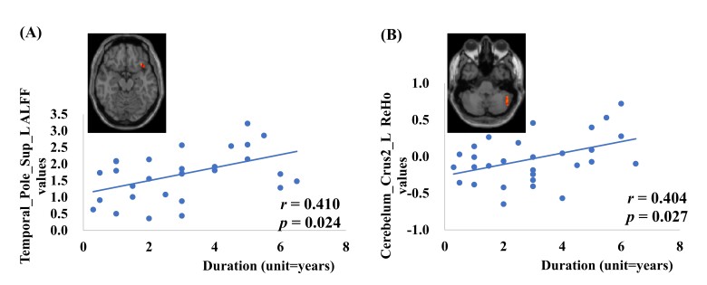
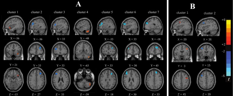
Results: Relative to HC, L_TN exhibited increased ALFF values in the left superior temporal gyrus and reduced values in the bilateral middle frontal gyrus. Elevated ReHo values were observed in the left cerebellar Crus2 region, while decreased values were identified in the bilateral middle frontal gyrus and left dorsolateral superior frontal gyrus. In R_TN, ALFF values increased in the left precentral gyrus and decreased in the right middle frontal gyrus; ReHo values remained unchanged. Correlation analysis indicated positive associations between disease duration and ALFF value of left superior temporal gyrus, as well as ReHo value of left cerebellar Crus2 region in L_TN.
Conclusions: This research indicated that both left and right TN patients exhibited changes in spontaneous brain activity during rest. These alterations predominantly occurred contralateral to the pain. These identified brain regions are implicated in pain perception, regulation, and emotional processing, suggesting their relevance to the modulation and adaptive changes of the human brain in response to trigeminal neuralgia.
期刊介绍:
Founded upon sound scientific principles, this journal continues to make important contributions that strongly influence the work of dental and medical professionals involved in treating oral and facial pain, including temporomandibular disorders, and headache. In addition to providing timely scientific research and clinical articles, the journal presents diagnostic techniques and treatment therapies for oral and facial pain, headache, mandibular dysfunction, and occlusion and covers pharmacology, physical therapy, surgery, and other pain-management methods.
 求助内容:
求助内容: 应助结果提醒方式:


