{"title":"辅助进行颞下颌关节镜手术的引导装置-初步研究。","authors":"Waseem Abboud, Shoshana Reiter, Pessia Friedman-Rubin, Dror Shamir, Oren Peleg","doi":"10.22514/jofph.2025.012","DOIUrl":null,"url":null,"abstract":"<p><strong>Background: </strong>Arthroscopic surgery of the temporomandibular joint (TMJ) requires inserting an arthroscope and a working cannula into the joint cavity. Working cannula introduction and positioning require high levels of expertise.</p><p><strong>Methods: </strong>A randomized controlled trial was performed on patients with closed lock of the TMJ who underwent arthroscopic lysis and lavage. A total of 15 subjects participated in this study, with 6 in the study group using the Locator-Positioner guide device (LOPO) and 9 in the control group using triangulation. The main outcomes included: (1) Number of attempts necessary for successful cannula insertion. (2) The time between arthroscope insertion and the appearance of the working cannula on the monitor, and (3) Overall surgery duration.</p><p><strong>Results: </strong>A successful cannula insertion took an average of 2.1 attempts in the study group compared with 3 attempts in the control group (<i>p</i> = 0.045). Study group arthroscope insertion to monitor appearance of cannula took 2.3 minutes, whereas control group took 4 minutes (<i>p</i> = 0.039). A total of 14 minutes was spent on surgery in the study group compared to 16.5 minutes in the control group (<i>p</i> = 0.009).</p><p><strong>Conclusions: </strong>LOPO device improved both the insertion of the working cannula into the TMJ and its positioning relative to the arthroscope throughout surgery. It reduced insertion attempts and shortened the surgery duration.</p><p><strong>Clinical trial registration: </strong>the study was registered at clinicaltrials.gov, identifier: NCT06520917.</p>","PeriodicalId":48800,"journal":{"name":"Journal of Oral & Facial Pain and Headache","volume":"39 1","pages":"128-133"},"PeriodicalIF":2.4000,"publicationDate":"2025-03-01","publicationTypes":"Journal Article","fieldsOfStudy":null,"isOpenAccess":false,"openAccessPdf":"https://www.ncbi.nlm.nih.gov/pmc/articles/PMC11934736/pdf/","citationCount":"0","resultStr":"{\"title\":\"Guide device to assist in performing arthroscopic surgery of the temporomandibular joint-a preliminary study.\",\"authors\":\"Waseem Abboud, Shoshana Reiter, Pessia Friedman-Rubin, Dror Shamir, Oren Peleg\",\"doi\":\"10.22514/jofph.2025.012\",\"DOIUrl\":null,\"url\":null,\"abstract\":\"<p><strong>Background: </strong>Arthroscopic surgery of the temporomandibular joint (TMJ) requires inserting an arthroscope and a working cannula into the joint cavity. Working cannula introduction and positioning require high levels of expertise.</p><p><strong>Methods: </strong>A randomized controlled trial was performed on patients with closed lock of the TMJ who underwent arthroscopic lysis and lavage. A total of 15 subjects participated in this study, with 6 in the study group using the Locator-Positioner guide device (LOPO) and 9 in the control group using triangulation. The main outcomes included: (1) Number of attempts necessary for successful cannula insertion. (2) The time between arthroscope insertion and the appearance of the working cannula on the monitor, and (3) Overall surgery duration.</p><p><strong>Results: </strong>A successful cannula insertion took an average of 2.1 attempts in the study group compared with 3 attempts in the control group (<i>p</i> = 0.045). Study group arthroscope insertion to monitor appearance of cannula took 2.3 minutes, whereas control group took 4 minutes (<i>p</i> = 0.039). A total of 14 minutes was spent on surgery in the study group compared to 16.5 minutes in the control group (<i>p</i> = 0.009).</p><p><strong>Conclusions: </strong>LOPO device improved both the insertion of the working cannula into the TMJ and its positioning relative to the arthroscope throughout surgery. It reduced insertion attempts and shortened the surgery duration.</p><p><strong>Clinical trial registration: </strong>the study was registered at clinicaltrials.gov, identifier: NCT06520917.</p>\",\"PeriodicalId\":48800,\"journal\":{\"name\":\"Journal of Oral & Facial Pain and Headache\",\"volume\":\"39 1\",\"pages\":\"128-133\"},\"PeriodicalIF\":2.4000,\"publicationDate\":\"2025-03-01\",\"publicationTypes\":\"Journal Article\",\"fieldsOfStudy\":null,\"isOpenAccess\":false,\"openAccessPdf\":\"https://www.ncbi.nlm.nih.gov/pmc/articles/PMC11934736/pdf/\",\"citationCount\":\"0\",\"resultStr\":null,\"platform\":\"Semanticscholar\",\"paperid\":null,\"PeriodicalName\":\"Journal of Oral & Facial Pain and Headache\",\"FirstCategoryId\":\"3\",\"ListUrlMain\":\"https://doi.org/10.22514/jofph.2025.012\",\"RegionNum\":3,\"RegionCategory\":\"医学\",\"ArticlePicture\":[],\"TitleCN\":null,\"AbstractTextCN\":null,\"PMCID\":null,\"EPubDate\":\"2025/3/12 0:00:00\",\"PubModel\":\"Epub\",\"JCR\":\"Q2\",\"JCRName\":\"DENTISTRY, ORAL SURGERY & MEDICINE\",\"Score\":null,\"Total\":0}","platform":"Semanticscholar","paperid":null,"PeriodicalName":"Journal of Oral & Facial Pain and Headache","FirstCategoryId":"3","ListUrlMain":"https://doi.org/10.22514/jofph.2025.012","RegionNum":3,"RegionCategory":"医学","ArticlePicture":[],"TitleCN":null,"AbstractTextCN":null,"PMCID":null,"EPubDate":"2025/3/12 0:00:00","PubModel":"Epub","JCR":"Q2","JCRName":"DENTISTRY, ORAL SURGERY & MEDICINE","Score":null,"Total":0}
引用次数: 0
摘要
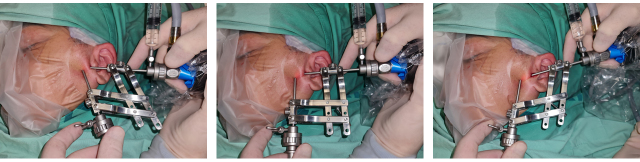
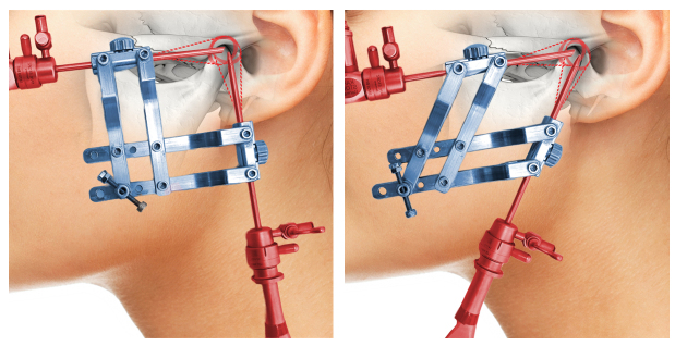
背景:颞下颌关节(TMJ)的关节镜手术需要在关节腔内插入关节镜和工作套管。工作套管的导入和定位需要高水平的专业知识。方法:对颞下颌关节闭锁患者行关节镜下松解灌洗进行随机对照试验。共15名受试者参与本研究,其中研究组6人使用定位器定位器(LOPO),对照组9人使用三角测量法。主要观察结果包括:(1)插管成功所需次数。(2)置入关节镜到工作套管出现在监护仪上的时间;(3)手术总时间。结果:研究组平均2.1次插管成功,对照组平均3次(p = 0.045)。研究组置入关节镜监测插管外观用时2.3 min,对照组置入关节镜监测插管外观用时4 min (p = 0.039)。研究组手术时间为14分钟,对照组为16.5分钟(p = 0.009)。结论:LOPO装置在整个手术过程中改善了工作套管插入TMJ和相对于关节镜的定位。它减少了插入次数,缩短了手术时间。临床试验注册:该研究已在clinicaltrials.gov注册,识别码:NCT06520917。
Guide device to assist in performing arthroscopic surgery of the temporomandibular joint-a preliminary study.
Background: Arthroscopic surgery of the temporomandibular joint (TMJ) requires inserting an arthroscope and a working cannula into the joint cavity. Working cannula introduction and positioning require high levels of expertise.
Methods: A randomized controlled trial was performed on patients with closed lock of the TMJ who underwent arthroscopic lysis and lavage. A total of 15 subjects participated in this study, with 6 in the study group using the Locator-Positioner guide device (LOPO) and 9 in the control group using triangulation. The main outcomes included: (1) Number of attempts necessary for successful cannula insertion. (2) The time between arthroscope insertion and the appearance of the working cannula on the monitor, and (3) Overall surgery duration.
Results: A successful cannula insertion took an average of 2.1 attempts in the study group compared with 3 attempts in the control group (p = 0.045). Study group arthroscope insertion to monitor appearance of cannula took 2.3 minutes, whereas control group took 4 minutes (p = 0.039). A total of 14 minutes was spent on surgery in the study group compared to 16.5 minutes in the control group (p = 0.009).
Conclusions: LOPO device improved both the insertion of the working cannula into the TMJ and its positioning relative to the arthroscope throughout surgery. It reduced insertion attempts and shortened the surgery duration.
Clinical trial registration: the study was registered at clinicaltrials.gov, identifier: NCT06520917.
期刊介绍:
Founded upon sound scientific principles, this journal continues to make important contributions that strongly influence the work of dental and medical professionals involved in treating oral and facial pain, including temporomandibular disorders, and headache. In addition to providing timely scientific research and clinical articles, the journal presents diagnostic techniques and treatment therapies for oral and facial pain, headache, mandibular dysfunction, and occlusion and covers pharmacology, physical therapy, surgery, and other pain-management methods.
 求助内容:
求助内容: 应助结果提醒方式:


