{"title":"HR-pQCT评估宫颈癌患者同步放化疗后骨微结构和体积矿物密度的动态变化:一项前瞻性研究。","authors":"Weishi Cheng, Yijun Wu, Jing Shen, Hui Guan, Li Zhang, Hongnan Zhen, Yinjie Tao, Weibo Xia, Zhikai Liu, Fuquan Zhang","doi":"10.1186/s40364-025-00754-6","DOIUrl":null,"url":null,"abstract":"<p><p>Bone changes in patients undergoing pelvic radiotherapy remain unclear. This study initially utilized high-resolution peripheral quantitative computed tomography (HR-pQCT) to assess the dynamic changes in bone microarchitecture and volumetric bone mineral density (BMD) in patients with cervical cancer before and after concurrent chemoradiotherapy. This prospective, observational study included patients with squamous carcinoma of the cervix scheduled for concurrent chemoradiotherapy. Patients underwent HR-pQCT, dual-energy X-ray absorptiometry (DXA) and laboratory tests before chemoradiotherapy, and at three and six months post-chemoradiotherapy. DXA, serving as the clinical standard for measuring BMD, was employed alongside HR-pQCT to provide complementary insights into bone micro-changes. The primary endpoint comprised changes in total (Tt.vBMD), trabecular (Tb.vBMD) and cortical (Ct.vBMD) volumetric BMD at the distal radius and tibia between pre-chemoradiotherapy and 6 months post-chemoradiotherapy. A total of 21 patients were enrolled, and one patient chose to withdraw (median age: 54.5 years). Tt.vBMD significantly decreased three months (distal radius: -1.65%, P = 0.008; distal tibia: -2.4%, P < 0.001) and six months (distal radius: -3.03%, P = 0.003; distal tibia: -2.69%, P = 0.002) post-chemoradiotherapy compared to baseline. Similarly, Tb.vBMD and Ct.vBMD demonstrated a significant downward trend post-chemoradiotherapy, with mean percent changes at three months of -0.73% and - 1.59% for the distal radius, and - 1.95% and - 1.50% for the distal tibia, respectively. The trends in BMD changes measured by DXA align with those observed using HR-pQCT. Regarding the laboratory tests, estradiol levels significantly decreased post-chemoradiotherapy, while follicle stimulating hormone and luteinizing hormone levels significantly increased. The results found that concurrent chemoradiotherapy was associated with the changes in bone volume, microstructure and BMD, especially in BMD three months post-chemoradiotherapy. Most of the bone micro-changes had not reverted by six months. This study explored the feasibility of early fracture risk identification post-chemoradiotherapy, aiding physicians in taking timely measures to improve prognosis.</p>","PeriodicalId":54225,"journal":{"name":"Biomarker Research","volume":"13 1","pages":"46"},"PeriodicalIF":11.5000,"publicationDate":"2025-03-18","publicationTypes":"Journal Article","fieldsOfStudy":null,"isOpenAccess":false,"openAccessPdf":"https://www.ncbi.nlm.nih.gov/pmc/articles/PMC11921580/pdf/","citationCount":"0","resultStr":"{\"title\":\"Dynamic changes of bone microarchitecture and volumetric mineral density assessed by HR-pQCT in patients with cervical cancer after concurrent chemoradiotherapy: a prospective study.\",\"authors\":\"Weishi Cheng, Yijun Wu, Jing Shen, Hui Guan, Li Zhang, Hongnan Zhen, Yinjie Tao, Weibo Xia, Zhikai Liu, Fuquan Zhang\",\"doi\":\"10.1186/s40364-025-00754-6\",\"DOIUrl\":null,\"url\":null,\"abstract\":\"<p><p>Bone changes in patients undergoing pelvic radiotherapy remain unclear. This study initially utilized high-resolution peripheral quantitative computed tomography (HR-pQCT) to assess the dynamic changes in bone microarchitecture and volumetric bone mineral density (BMD) in patients with cervical cancer before and after concurrent chemoradiotherapy. This prospective, observational study included patients with squamous carcinoma of the cervix scheduled for concurrent chemoradiotherapy. Patients underwent HR-pQCT, dual-energy X-ray absorptiometry (DXA) and laboratory tests before chemoradiotherapy, and at three and six months post-chemoradiotherapy. DXA, serving as the clinical standard for measuring BMD, was employed alongside HR-pQCT to provide complementary insights into bone micro-changes. The primary endpoint comprised changes in total (Tt.vBMD), trabecular (Tb.vBMD) and cortical (Ct.vBMD) volumetric BMD at the distal radius and tibia between pre-chemoradiotherapy and 6 months post-chemoradiotherapy. A total of 21 patients were enrolled, and one patient chose to withdraw (median age: 54.5 years). Tt.vBMD significantly decreased three months (distal radius: -1.65%, P = 0.008; distal tibia: -2.4%, P < 0.001) and six months (distal radius: -3.03%, P = 0.003; distal tibia: -2.69%, P = 0.002) post-chemoradiotherapy compared to baseline. Similarly, Tb.vBMD and Ct.vBMD demonstrated a significant downward trend post-chemoradiotherapy, with mean percent changes at three months of -0.73% and - 1.59% for the distal radius, and - 1.95% and - 1.50% for the distal tibia, respectively. The trends in BMD changes measured by DXA align with those observed using HR-pQCT. Regarding the laboratory tests, estradiol levels significantly decreased post-chemoradiotherapy, while follicle stimulating hormone and luteinizing hormone levels significantly increased. The results found that concurrent chemoradiotherapy was associated with the changes in bone volume, microstructure and BMD, especially in BMD three months post-chemoradiotherapy. Most of the bone micro-changes had not reverted by six months. This study explored the feasibility of early fracture risk identification post-chemoradiotherapy, aiding physicians in taking timely measures to improve prognosis.</p>\",\"PeriodicalId\":54225,\"journal\":{\"name\":\"Biomarker Research\",\"volume\":\"13 1\",\"pages\":\"46\"},\"PeriodicalIF\":11.5000,\"publicationDate\":\"2025-03-18\",\"publicationTypes\":\"Journal Article\",\"fieldsOfStudy\":null,\"isOpenAccess\":false,\"openAccessPdf\":\"https://www.ncbi.nlm.nih.gov/pmc/articles/PMC11921580/pdf/\",\"citationCount\":\"0\",\"resultStr\":null,\"platform\":\"Semanticscholar\",\"paperid\":null,\"PeriodicalName\":\"Biomarker Research\",\"FirstCategoryId\":\"3\",\"ListUrlMain\":\"https://doi.org/10.1186/s40364-025-00754-6\",\"RegionNum\":2,\"RegionCategory\":\"医学\",\"ArticlePicture\":[],\"TitleCN\":null,\"AbstractTextCN\":null,\"PMCID\":null,\"EPubDate\":\"\",\"PubModel\":\"\",\"JCR\":\"Q1\",\"JCRName\":\"MEDICINE, RESEARCH & EXPERIMENTAL\",\"Score\":null,\"Total\":0}","platform":"Semanticscholar","paperid":null,"PeriodicalName":"Biomarker Research","FirstCategoryId":"3","ListUrlMain":"https://doi.org/10.1186/s40364-025-00754-6","RegionNum":2,"RegionCategory":"医学","ArticlePicture":[],"TitleCN":null,"AbstractTextCN":null,"PMCID":null,"EPubDate":"","PubModel":"","JCR":"Q1","JCRName":"MEDICINE, RESEARCH & EXPERIMENTAL","Score":null,"Total":0}
Dynamic changes of bone microarchitecture and volumetric mineral density assessed by HR-pQCT in patients with cervical cancer after concurrent chemoradiotherapy: a prospective study.
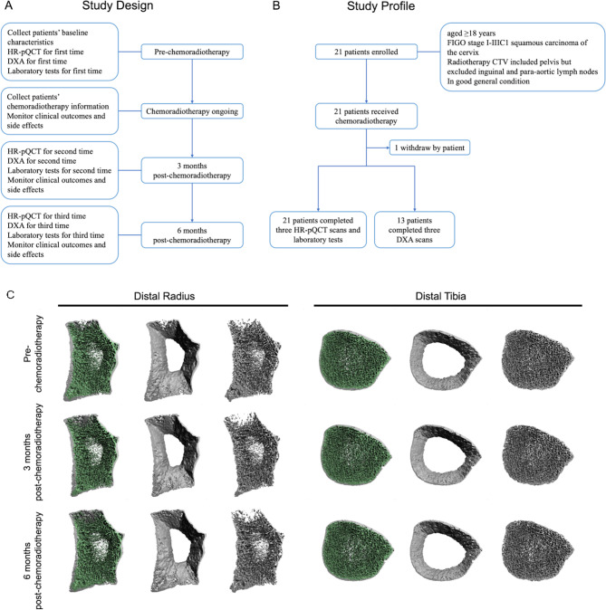
Bone changes in patients undergoing pelvic radiotherapy remain unclear. This study initially utilized high-resolution peripheral quantitative computed tomography (HR-pQCT) to assess the dynamic changes in bone microarchitecture and volumetric bone mineral density (BMD) in patients with cervical cancer before and after concurrent chemoradiotherapy. This prospective, observational study included patients with squamous carcinoma of the cervix scheduled for concurrent chemoradiotherapy. Patients underwent HR-pQCT, dual-energy X-ray absorptiometry (DXA) and laboratory tests before chemoradiotherapy, and at three and six months post-chemoradiotherapy. DXA, serving as the clinical standard for measuring BMD, was employed alongside HR-pQCT to provide complementary insights into bone micro-changes. The primary endpoint comprised changes in total (Tt.vBMD), trabecular (Tb.vBMD) and cortical (Ct.vBMD) volumetric BMD at the distal radius and tibia between pre-chemoradiotherapy and 6 months post-chemoradiotherapy. A total of 21 patients were enrolled, and one patient chose to withdraw (median age: 54.5 years). Tt.vBMD significantly decreased three months (distal radius: -1.65%, P = 0.008; distal tibia: -2.4%, P < 0.001) and six months (distal radius: -3.03%, P = 0.003; distal tibia: -2.69%, P = 0.002) post-chemoradiotherapy compared to baseline. Similarly, Tb.vBMD and Ct.vBMD demonstrated a significant downward trend post-chemoradiotherapy, with mean percent changes at three months of -0.73% and - 1.59% for the distal radius, and - 1.95% and - 1.50% for the distal tibia, respectively. The trends in BMD changes measured by DXA align with those observed using HR-pQCT. Regarding the laboratory tests, estradiol levels significantly decreased post-chemoradiotherapy, while follicle stimulating hormone and luteinizing hormone levels significantly increased. The results found that concurrent chemoradiotherapy was associated with the changes in bone volume, microstructure and BMD, especially in BMD three months post-chemoradiotherapy. Most of the bone micro-changes had not reverted by six months. This study explored the feasibility of early fracture risk identification post-chemoradiotherapy, aiding physicians in taking timely measures to improve prognosis.
Biomarker ResearchBiochemistry, Genetics and Molecular Biology-Molecular Medicine
CiteScore
15.80
自引率
1.80%
发文量
80
审稿时长
10 weeks
期刊介绍:
Biomarker Research, an open-access, peer-reviewed journal, covers all aspects of biomarker investigation. It seeks to publish original discoveries, novel concepts, commentaries, and reviews across various biomedical disciplines. The field of biomarker research has progressed significantly with the rise of personalized medicine and individual health. Biomarkers play a crucial role in drug discovery and development, as well as in disease diagnosis, treatment, prognosis, and prevention, particularly in the genome era.

 求助内容:
求助内容: 应助结果提醒方式:


