{"title":"光子计数计算机断层扫描在儿童心血管成像中的作用。","authors":"Arosh S Perera Molligoda Arachchige, Yash Verma","doi":"10.5409/wjcp.v14.i1.99288","DOIUrl":null,"url":null,"abstract":"<p><p>Photon-counting computed tomography (PCCT) represents a significant advancement in pediatric cardiovascular imaging. Traditional CT systems employ energy-integrating detectors that convert X-ray photons into visible light, whereas PCCT utilizes photon-counting detectors that directly transform X-ray photons into electric signals. This direct conversion allows photon-counting detectors to sort photons into discrete energy levels, thereby enhancing image quality through superior noise reduction, improved spatial and contrast resolution, and reduced artifacts. In pediatric applications, PCCT offers substantial benefits, including lower radiation doses, which may help reduce the risk of malignancy in pediatric patients, with perhaps greater potential to benefit those with repeated exposure from a young age. Enhanced spatial resolution facilitates better visualization of small structures, vital for diagnosing congenital heart defects. Additionally, PCCT's spectral capabilities improve tissue characterization and enable the creation of virtual monoenergetic images, which enhance soft-tissue contrast and potentially reduce contrast media doses. Initial clinical results indicate that PCCT provides superior image quality and diagnostic accuracy compared to conventional CT, particularly in challenging pediatric cardiovascular cases. As PCCT technology matures, further research and standardized protocols will be essential to fully integrate it into pediatric imaging practices, ensuring optimized diagnostic outcomes and patient safety.</p>","PeriodicalId":75338,"journal":{"name":"World journal of clinical pediatrics","volume":"14 1","pages":"99288"},"PeriodicalIF":0.0000,"publicationDate":"2025-03-09","publicationTypes":"Journal Article","fieldsOfStudy":null,"isOpenAccess":false,"openAccessPdf":"https://www.ncbi.nlm.nih.gov/pmc/articles/PMC11686577/pdf/","citationCount":"0","resultStr":"{\"title\":\"Role of photon-counting computed tomography in pediatric cardiovascular imaging.\",\"authors\":\"Arosh S Perera Molligoda Arachchige, Yash Verma\",\"doi\":\"10.5409/wjcp.v14.i1.99288\",\"DOIUrl\":null,\"url\":null,\"abstract\":\"<p><p>Photon-counting computed tomography (PCCT) represents a significant advancement in pediatric cardiovascular imaging. Traditional CT systems employ energy-integrating detectors that convert X-ray photons into visible light, whereas PCCT utilizes photon-counting detectors that directly transform X-ray photons into electric signals. This direct conversion allows photon-counting detectors to sort photons into discrete energy levels, thereby enhancing image quality through superior noise reduction, improved spatial and contrast resolution, and reduced artifacts. In pediatric applications, PCCT offers substantial benefits, including lower radiation doses, which may help reduce the risk of malignancy in pediatric patients, with perhaps greater potential to benefit those with repeated exposure from a young age. Enhanced spatial resolution facilitates better visualization of small structures, vital for diagnosing congenital heart defects. Additionally, PCCT's spectral capabilities improve tissue characterization and enable the creation of virtual monoenergetic images, which enhance soft-tissue contrast and potentially reduce contrast media doses. Initial clinical results indicate that PCCT provides superior image quality and diagnostic accuracy compared to conventional CT, particularly in challenging pediatric cardiovascular cases. As PCCT technology matures, further research and standardized protocols will be essential to fully integrate it into pediatric imaging practices, ensuring optimized diagnostic outcomes and patient safety.</p>\",\"PeriodicalId\":75338,\"journal\":{\"name\":\"World journal of clinical pediatrics\",\"volume\":\"14 1\",\"pages\":\"99288\"},\"PeriodicalIF\":0.0000,\"publicationDate\":\"2025-03-09\",\"publicationTypes\":\"Journal Article\",\"fieldsOfStudy\":null,\"isOpenAccess\":false,\"openAccessPdf\":\"https://www.ncbi.nlm.nih.gov/pmc/articles/PMC11686577/pdf/\",\"citationCount\":\"0\",\"resultStr\":null,\"platform\":\"Semanticscholar\",\"paperid\":null,\"PeriodicalName\":\"World journal of clinical pediatrics\",\"FirstCategoryId\":\"1085\",\"ListUrlMain\":\"https://doi.org/10.5409/wjcp.v14.i1.99288\",\"RegionNum\":0,\"RegionCategory\":null,\"ArticlePicture\":[],\"TitleCN\":null,\"AbstractTextCN\":null,\"PMCID\":null,\"EPubDate\":\"\",\"PubModel\":\"\",\"JCR\":\"\",\"JCRName\":\"\",\"Score\":null,\"Total\":0}","platform":"Semanticscholar","paperid":null,"PeriodicalName":"World journal of clinical pediatrics","FirstCategoryId":"1085","ListUrlMain":"https://doi.org/10.5409/wjcp.v14.i1.99288","RegionNum":0,"RegionCategory":null,"ArticlePicture":[],"TitleCN":null,"AbstractTextCN":null,"PMCID":null,"EPubDate":"","PubModel":"","JCR":"","JCRName":"","Score":null,"Total":0}
Role of photon-counting computed tomography in pediatric cardiovascular imaging.
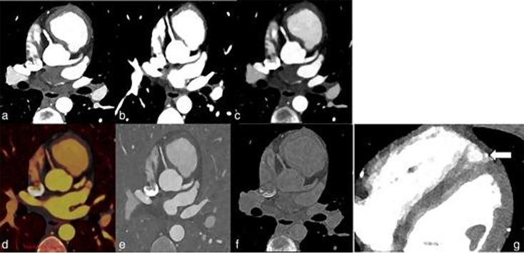
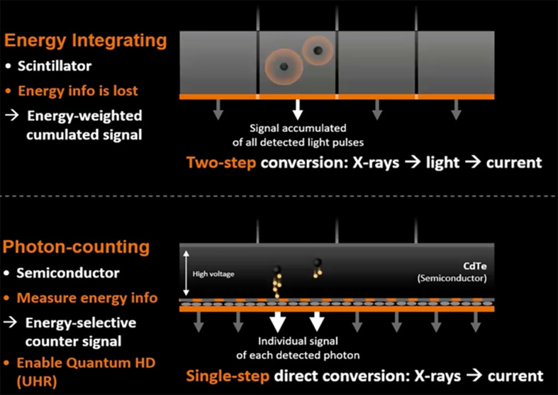
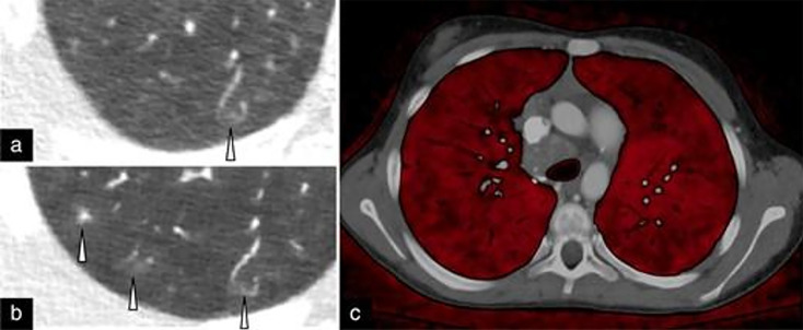
Photon-counting computed tomography (PCCT) represents a significant advancement in pediatric cardiovascular imaging. Traditional CT systems employ energy-integrating detectors that convert X-ray photons into visible light, whereas PCCT utilizes photon-counting detectors that directly transform X-ray photons into electric signals. This direct conversion allows photon-counting detectors to sort photons into discrete energy levels, thereby enhancing image quality through superior noise reduction, improved spatial and contrast resolution, and reduced artifacts. In pediatric applications, PCCT offers substantial benefits, including lower radiation doses, which may help reduce the risk of malignancy in pediatric patients, with perhaps greater potential to benefit those with repeated exposure from a young age. Enhanced spatial resolution facilitates better visualization of small structures, vital for diagnosing congenital heart defects. Additionally, PCCT's spectral capabilities improve tissue characterization and enable the creation of virtual monoenergetic images, which enhance soft-tissue contrast and potentially reduce contrast media doses. Initial clinical results indicate that PCCT provides superior image quality and diagnostic accuracy compared to conventional CT, particularly in challenging pediatric cardiovascular cases. As PCCT technology matures, further research and standardized protocols will be essential to fully integrate it into pediatric imaging practices, ensuring optimized diagnostic outcomes and patient safety.

 求助内容:
求助内容: 应助结果提醒方式:


