Jakob Ackermann, Berfin Caliskan, Martin Hartmann, Lazaros Vlachopoulos, Sandro F Fucentese
{"title":"滑车发育不良患者滑车上骨刺对髌股软骨的影响。","authors":"Jakob Ackermann, Berfin Caliskan, Martin Hartmann, Lazaros Vlachopoulos, Sandro F Fucentese","doi":"10.1177/03635465251323806","DOIUrl":null,"url":null,"abstract":"<p><strong>Background: </strong>The presence of a supratrochlear spur has been shown to influence outcomes in patients with trochlear dysplasia and is thought to accelerate cartilage wear. However, the current literature does not provide an evidence-based threshold for a relevant supratrochlear spur height.</p><p><strong>Purpose/hypothesis: </strong>The purpose of this study was to establish a clinically significant supratrochlear spur height associated with patellofemoral chondral damage to guide surgeons in surgical decision-making. It was hypothesized that a supratrochlear spur negatively affects patellofemoral articular cartilage, with large spurs having the greatest effect.</p><p><strong>Study design: </strong>Case series; Level of evidence, 4.</p><p><strong>Methods: </strong>This study evaluated 363 knees with trochlear dysplasia that were scheduled to undergo surgery for the treatment of patellar instability at a single institution. All patients underwent preoperative true lateral radiography and magnetic resonance imaging (MRI). There were 2 independent reviewers who analyzed the supratrochlear spur height by measuring the distance between a tangent at the anterior femoral cortex and the most prominent point of the trochlea on sagittal MRI as well as other common patellofemoral parameters. All MRI scans were assessed for full-thickness cartilage lesions.</p><p><strong>Results: </strong>Of the included 363 knees, 91 (25.1%) showed full-thickness cartilage defects on the patella, while 21 (5.8%) had full-thickness trochlear cartilage damage. Patellar defects were significantly correlated with patient's age (<i>r</i> = 0.237; <i>P</i> < .001), body mass index (<i>r</i> = 0.148; <i>P</i> = .005), and supratrochlear spur height (<i>r</i> = 0.196; <i>P</i> < .001). Trochlear defects were significantly associated with patient's age (<i>r</i> = 0.160; <i>P</i> = .002), patellar tilt (<i>r</i> = 0.202; <i>P</i> < .001), tibial tubercle-trochlear groove distance (<i>r</i> = 0.128; <i>P</i> = .014), and supratrochlear spur height (<i>r</i> = 0.151; <i>P</i> < .004). Trochlear dysplasia types B and D showed a trend toward a higher prevalence in patellar defects (<i>P</i> = .082), while they were significantly associated with a higher prevalance of trochlear defects (<i>P</i> = .003) compared with types A and C. Knees with patellar (5.1 ± 2.0 vs 4.3 ± 1.7 mm, respectively; <i>P</i> = .001) and trochlear (5.3 ± 2.1 vs 4.4 ± 1.8 mm, respectively; <i>P</i> = .015) cartilage defects had a significantly larger supratrochlear spur height than knees without patellar and trochlear defects. A supratrochlear spur height ≥6 mm had adjusted odds ratios of 2.7 (95% CI, 1.6-4.5; <i>P</i> < .001) and 3.4 (95% CI, 1.3-8.8; <i>P</i> = .014) for developing patellar and trochlear cartilage damage, respectively.</p><p><strong>Conclusion: </strong>A supratrochlear spur was significantly associated with patellofemoral cartilage damage. Large supratrochlear spurs demonstrated a substantially increased risk of developing patellofemoral cartilage damage.</p>","PeriodicalId":55528,"journal":{"name":"American Journal of Sports Medicine","volume":" ","pages":"1127-1132"},"PeriodicalIF":4.5000,"publicationDate":"2025-04-01","publicationTypes":"Journal Article","fieldsOfStudy":null,"isOpenAccess":false,"openAccessPdf":"https://www.ncbi.nlm.nih.gov/pmc/articles/PMC11951347/pdf/","citationCount":"0","resultStr":"{\"title\":\"The Effect of a Supratrochlear Spur on Patellofemoral Cartilage in Patients With Trochlear Dysplasia.\",\"authors\":\"Jakob Ackermann, Berfin Caliskan, Martin Hartmann, Lazaros Vlachopoulos, Sandro F Fucentese\",\"doi\":\"10.1177/03635465251323806\",\"DOIUrl\":null,\"url\":null,\"abstract\":\"<p><strong>Background: </strong>The presence of a supratrochlear spur has been shown to influence outcomes in patients with trochlear dysplasia and is thought to accelerate cartilage wear. However, the current literature does not provide an evidence-based threshold for a relevant supratrochlear spur height.</p><p><strong>Purpose/hypothesis: </strong>The purpose of this study was to establish a clinically significant supratrochlear spur height associated with patellofemoral chondral damage to guide surgeons in surgical decision-making. It was hypothesized that a supratrochlear spur negatively affects patellofemoral articular cartilage, with large spurs having the greatest effect.</p><p><strong>Study design: </strong>Case series; Level of evidence, 4.</p><p><strong>Methods: </strong>This study evaluated 363 knees with trochlear dysplasia that were scheduled to undergo surgery for the treatment of patellar instability at a single institution. All patients underwent preoperative true lateral radiography and magnetic resonance imaging (MRI). There were 2 independent reviewers who analyzed the supratrochlear spur height by measuring the distance between a tangent at the anterior femoral cortex and the most prominent point of the trochlea on sagittal MRI as well as other common patellofemoral parameters. All MRI scans were assessed for full-thickness cartilage lesions.</p><p><strong>Results: </strong>Of the included 363 knees, 91 (25.1%) showed full-thickness cartilage defects on the patella, while 21 (5.8%) had full-thickness trochlear cartilage damage. Patellar defects were significantly correlated with patient's age (<i>r</i> = 0.237; <i>P</i> < .001), body mass index (<i>r</i> = 0.148; <i>P</i> = .005), and supratrochlear spur height (<i>r</i> = 0.196; <i>P</i> < .001). Trochlear defects were significantly associated with patient's age (<i>r</i> = 0.160; <i>P</i> = .002), patellar tilt (<i>r</i> = 0.202; <i>P</i> < .001), tibial tubercle-trochlear groove distance (<i>r</i> = 0.128; <i>P</i> = .014), and supratrochlear spur height (<i>r</i> = 0.151; <i>P</i> < .004). Trochlear dysplasia types B and D showed a trend toward a higher prevalence in patellar defects (<i>P</i> = .082), while they were significantly associated with a higher prevalance of trochlear defects (<i>P</i> = .003) compared with types A and C. Knees with patellar (5.1 ± 2.0 vs 4.3 ± 1.7 mm, respectively; <i>P</i> = .001) and trochlear (5.3 ± 2.1 vs 4.4 ± 1.8 mm, respectively; <i>P</i> = .015) cartilage defects had a significantly larger supratrochlear spur height than knees without patellar and trochlear defects. A supratrochlear spur height ≥6 mm had adjusted odds ratios of 2.7 (95% CI, 1.6-4.5; <i>P</i> < .001) and 3.4 (95% CI, 1.3-8.8; <i>P</i> = .014) for developing patellar and trochlear cartilage damage, respectively.</p><p><strong>Conclusion: </strong>A supratrochlear spur was significantly associated with patellofemoral cartilage damage. Large supratrochlear spurs demonstrated a substantially increased risk of developing patellofemoral cartilage damage.</p>\",\"PeriodicalId\":55528,\"journal\":{\"name\":\"American Journal of Sports Medicine\",\"volume\":\" \",\"pages\":\"1127-1132\"},\"PeriodicalIF\":4.5000,\"publicationDate\":\"2025-04-01\",\"publicationTypes\":\"Journal Article\",\"fieldsOfStudy\":null,\"isOpenAccess\":false,\"openAccessPdf\":\"https://www.ncbi.nlm.nih.gov/pmc/articles/PMC11951347/pdf/\",\"citationCount\":\"0\",\"resultStr\":null,\"platform\":\"Semanticscholar\",\"paperid\":null,\"PeriodicalName\":\"American Journal of Sports Medicine\",\"FirstCategoryId\":\"3\",\"ListUrlMain\":\"https://doi.org/10.1177/03635465251323806\",\"RegionNum\":1,\"RegionCategory\":\"医学\",\"ArticlePicture\":[],\"TitleCN\":null,\"AbstractTextCN\":null,\"PMCID\":null,\"EPubDate\":\"2025/3/4 0:00:00\",\"PubModel\":\"Epub\",\"JCR\":\"Q1\",\"JCRName\":\"ORTHOPEDICS\",\"Score\":null,\"Total\":0}","platform":"Semanticscholar","paperid":null,"PeriodicalName":"American Journal of Sports Medicine","FirstCategoryId":"3","ListUrlMain":"https://doi.org/10.1177/03635465251323806","RegionNum":1,"RegionCategory":"医学","ArticlePicture":[],"TitleCN":null,"AbstractTextCN":null,"PMCID":null,"EPubDate":"2025/3/4 0:00:00","PubModel":"Epub","JCR":"Q1","JCRName":"ORTHOPEDICS","Score":null,"Total":0}
引用次数: 0
摘要
背景:蜗上距的存在已被证明会影响蜗轮发育不良患者的预后,并被认为会加速软骨磨损。然而,目前的文献并未提供相关弧上骨刺高度的循证阈值:本研究的目的是确定与髌骨软骨损伤相关的具有临床意义的髌上距高度,以指导外科医生做出手术决策。假设髌骨上骨刺会对髌骨关节软骨产生负面影响,而大的骨刺影响最大:研究设计:病例系列;证据级别,4.方法:本研究评估了一家医疗机构中363例因髌骨不稳而计划接受手术治疗的髌骨发育不良膝关节。所有患者均接受了术前真侧位X光检查和磁共振成像(MRI)检查。两名独立审查员通过测量股骨前皮质切线与矢状磁共振成像上髌骨最突出点之间的距离来分析髌骨上距高度,并分析其他常见的髌股关节参数。所有核磁共振扫描均评估了全厚软骨病变:结果:在363个膝关节中,91个(25.1%)膝关节的髌骨软骨全厚缺损,21个(5.8%)膝关节的髌骨软骨全厚损伤。髌骨缺损与患者的年龄(r = 0.237; P < .001)、体重指数(r = 0.148; P = .005)和髌上距高度(r = 0.196; P < .001)有明显相关性。蝶骨缺损与患者的年龄(r = 0.160; P = .002)、髌骨倾斜度(r = 0.202; P < .001)、胫骨结节-蝶骨沟距离(r = 0.128; P = .014)和蝶骨上距高度(r = 0.151; P < .004)明显相关。与 A 型和 C 型相比,B 型和 D 型髌骨发育不良与较高的髌骨缺损患病率(P = .082)呈显著相关(P = .003)。1 ± 2.0 vs 4.3 ± 1.7 mm,P = .001)和喙突(5.3 ± 2.1 vs 4.4 ± 1.8 mm,P = .015)软骨缺损的膝关节的喙突上距高度明显大于无髌骨和喙突缺损的膝关节。髌上距高度≥6毫米的膝关节发生髌骨和蹄骨软骨损伤的调整后几率分别为2.7(95% CI,1.6-4.5;P < .001)和3.4(95% CI,1.3-8.8;P = .014):结论:髌骨髁上骨刺与髌骨软骨损伤密切相关。结论:髌骨上骨刺与髌骨软骨损伤密切相关,大的髌骨上骨刺会大大增加髌骨软骨损伤的风险。
The Effect of a Supratrochlear Spur on Patellofemoral Cartilage in Patients With Trochlear Dysplasia.
Background: The presence of a supratrochlear spur has been shown to influence outcomes in patients with trochlear dysplasia and is thought to accelerate cartilage wear. However, the current literature does not provide an evidence-based threshold for a relevant supratrochlear spur height.
Purpose/hypothesis: The purpose of this study was to establish a clinically significant supratrochlear spur height associated with patellofemoral chondral damage to guide surgeons in surgical decision-making. It was hypothesized that a supratrochlear spur negatively affects patellofemoral articular cartilage, with large spurs having the greatest effect.
Study design: Case series; Level of evidence, 4.
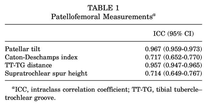
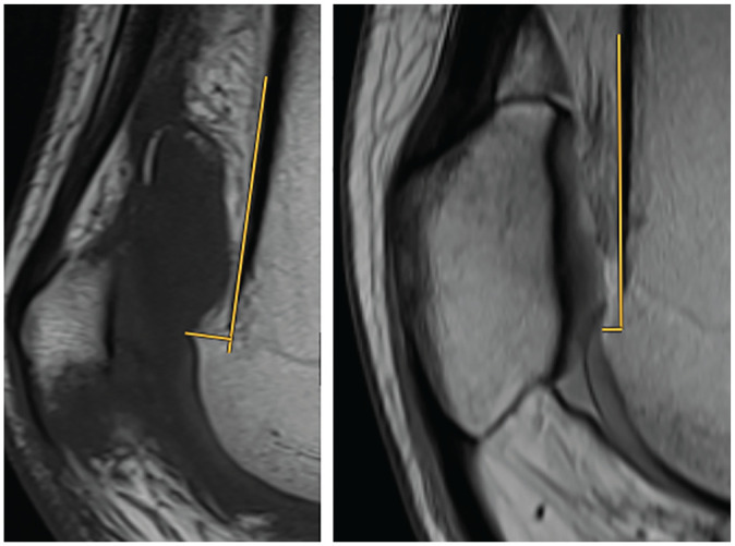
Methods: This study evaluated 363 knees with trochlear dysplasia that were scheduled to undergo surgery for the treatment of patellar instability at a single institution. All patients underwent preoperative true lateral radiography and magnetic resonance imaging (MRI). There were 2 independent reviewers who analyzed the supratrochlear spur height by measuring the distance between a tangent at the anterior femoral cortex and the most prominent point of the trochlea on sagittal MRI as well as other common patellofemoral parameters. All MRI scans were assessed for full-thickness cartilage lesions.
Results: Of the included 363 knees, 91 (25.1%) showed full-thickness cartilage defects on the patella, while 21 (5.8%) had full-thickness trochlear cartilage damage. Patellar defects were significantly correlated with patient's age (r = 0.237; P < .001), body mass index (r = 0.148; P = .005), and supratrochlear spur height (r = 0.196; P < .001). Trochlear defects were significantly associated with patient's age (r = 0.160; P = .002), patellar tilt (r = 0.202; P < .001), tibial tubercle-trochlear groove distance (r = 0.128; P = .014), and supratrochlear spur height (r = 0.151; P < .004). Trochlear dysplasia types B and D showed a trend toward a higher prevalence in patellar defects (P = .082), while they were significantly associated with a higher prevalance of trochlear defects (P = .003) compared with types A and C. Knees with patellar (5.1 ± 2.0 vs 4.3 ± 1.7 mm, respectively; P = .001) and trochlear (5.3 ± 2.1 vs 4.4 ± 1.8 mm, respectively; P = .015) cartilage defects had a significantly larger supratrochlear spur height than knees without patellar and trochlear defects. A supratrochlear spur height ≥6 mm had adjusted odds ratios of 2.7 (95% CI, 1.6-4.5; P < .001) and 3.4 (95% CI, 1.3-8.8; P = .014) for developing patellar and trochlear cartilage damage, respectively.
Conclusion: A supratrochlear spur was significantly associated with patellofemoral cartilage damage. Large supratrochlear spurs demonstrated a substantially increased risk of developing patellofemoral cartilage damage.
期刊介绍:
An invaluable resource for the orthopaedic sports medicine community, _The American Journal of Sports Medicine_ is a peer-reviewed scientific journal, first published in 1972. It is the official publication of the [American Orthopaedic Society for Sports Medicine (AOSSM)](http://www.sportsmed.org/)! The journal acts as an important forum for independent orthopaedic sports medicine research and education, allowing clinical practitioners the ability to make decisions based on sound scientific information.
This journal is a must-read for:
* Orthopaedic Surgeons and Specialists
* Sports Medicine Physicians
* Physiatrists
* Athletic Trainers
* Team Physicians
* And Physical Therapists

 求助内容:
求助内容: 应助结果提醒方式:


