Aglae Velasco Gonzalez, Liu Jingyu, Boris Buerke, Dennis Görlich, Joaquin Ortega-Quintanilla, Cristina Sauerland, Norbert Meier, Walter Heindel
{"title":"灌注模式作为紧急卒中诊断的工具:区分近端和远端MCA闭塞。","authors":"Aglae Velasco Gonzalez, Liu Jingyu, Boris Buerke, Dennis Görlich, Joaquin Ortega-Quintanilla, Cristina Sauerland, Norbert Meier, Walter Heindel","doi":"10.1136/bmjno-2024-001001","DOIUrl":null,"url":null,"abstract":"<p><strong>Background: </strong>To evaluate the effectiveness of a novel Perfusion Pattern (PP) scale in differentiating between proximal and distal middle cerebral artery (MCA) occlusions in patients with acute ischaemic stroke.</p><p><strong>Methods: </strong>This retrospective study included 201 patients with acute ischaemic stroke, categorised into two groups: those with M1 segment occlusions (n=114) and those with distal medium vessel occlusions (n=87). We analysed multimodal stroke CT imaging and clinical data, focusing on the occlusion site, hypoperfusion extent and basal ganglia involvement. Patients with tandem stenosis or multiple acute occlusions were excluded. Perfusion patterns were categorised into three types (PP-1, PP-2 and PP-3) based on the extent of hypoperfusion. Statistical analysis explored associations between the occlusion site, perfusion pattern and collateral status.</p><p><strong>Results: </strong>Among the 201 patients (mean age 75±14 years, 86 men), PP-1 was observed in 36.8% of patients (74/201), PP-2 in 27.4% (55/201) and PP-3 in 35.8% (72/201). The distribution of PP varied significantly by occlusion site (p<0.0001). Distal medium vessel occlusions were associated with PP-1 in 78.4% of cases (58/74), while PP-3 was most prevalent in M1 occlusions (90.3%, 65/72). The contingency coefficient revealed that occlusion location had a stronger association with the perfusion pattern (c=0.556) than collateral type (c=0.245). However, 21.6% of M1 occlusions (16/74) showed a PP-1 pattern and 9.7% of distal medium vessel occlusions (7/72) exhibited PP-3. Basal ganglia infarction presence was a reliable indicator of M1 occlusion with a 94% likelihood.</p><p><strong>Conclusions: </strong>Perfusion patterns can effectively differentiate between proximal and distal medium vessel MCA occlusions, aiding targeted assessment of CT angiography.</p>","PeriodicalId":52754,"journal":{"name":"BMJ Neurology Open","volume":"7 1","pages":"e001001"},"PeriodicalIF":2.4000,"publicationDate":"2025-02-27","publicationTypes":"Journal Article","fieldsOfStudy":null,"isOpenAccess":false,"openAccessPdf":"https://www.ncbi.nlm.nih.gov/pmc/articles/PMC11873334/pdf/","citationCount":"0","resultStr":"{\"title\":\"Perfusion patterns as a tool for emergency stroke diagnosis: differentiating proximal and distal MCA occlusions.\",\"authors\":\"Aglae Velasco Gonzalez, Liu Jingyu, Boris Buerke, Dennis Görlich, Joaquin Ortega-Quintanilla, Cristina Sauerland, Norbert Meier, Walter Heindel\",\"doi\":\"10.1136/bmjno-2024-001001\",\"DOIUrl\":null,\"url\":null,\"abstract\":\"<p><strong>Background: </strong>To evaluate the effectiveness of a novel Perfusion Pattern (PP) scale in differentiating between proximal and distal middle cerebral artery (MCA) occlusions in patients with acute ischaemic stroke.</p><p><strong>Methods: </strong>This retrospective study included 201 patients with acute ischaemic stroke, categorised into two groups: those with M1 segment occlusions (n=114) and those with distal medium vessel occlusions (n=87). We analysed multimodal stroke CT imaging and clinical data, focusing on the occlusion site, hypoperfusion extent and basal ganglia involvement. Patients with tandem stenosis or multiple acute occlusions were excluded. Perfusion patterns were categorised into three types (PP-1, PP-2 and PP-3) based on the extent of hypoperfusion. Statistical analysis explored associations between the occlusion site, perfusion pattern and collateral status.</p><p><strong>Results: </strong>Among the 201 patients (mean age 75±14 years, 86 men), PP-1 was observed in 36.8% of patients (74/201), PP-2 in 27.4% (55/201) and PP-3 in 35.8% (72/201). The distribution of PP varied significantly by occlusion site (p<0.0001). Distal medium vessel occlusions were associated with PP-1 in 78.4% of cases (58/74), while PP-3 was most prevalent in M1 occlusions (90.3%, 65/72). The contingency coefficient revealed that occlusion location had a stronger association with the perfusion pattern (c=0.556) than collateral type (c=0.245). However, 21.6% of M1 occlusions (16/74) showed a PP-1 pattern and 9.7% of distal medium vessel occlusions (7/72) exhibited PP-3. Basal ganglia infarction presence was a reliable indicator of M1 occlusion with a 94% likelihood.</p><p><strong>Conclusions: </strong>Perfusion patterns can effectively differentiate between proximal and distal medium vessel MCA occlusions, aiding targeted assessment of CT angiography.</p>\",\"PeriodicalId\":52754,\"journal\":{\"name\":\"BMJ Neurology Open\",\"volume\":\"7 1\",\"pages\":\"e001001\"},\"PeriodicalIF\":2.4000,\"publicationDate\":\"2025-02-27\",\"publicationTypes\":\"Journal Article\",\"fieldsOfStudy\":null,\"isOpenAccess\":false,\"openAccessPdf\":\"https://www.ncbi.nlm.nih.gov/pmc/articles/PMC11873334/pdf/\",\"citationCount\":\"0\",\"resultStr\":null,\"platform\":\"Semanticscholar\",\"paperid\":null,\"PeriodicalName\":\"BMJ Neurology Open\",\"FirstCategoryId\":\"1085\",\"ListUrlMain\":\"https://doi.org/10.1136/bmjno-2024-001001\",\"RegionNum\":0,\"RegionCategory\":null,\"ArticlePicture\":[],\"TitleCN\":null,\"AbstractTextCN\":null,\"PMCID\":null,\"EPubDate\":\"2025/1/1 0:00:00\",\"PubModel\":\"eCollection\",\"JCR\":\"Q3\",\"JCRName\":\"CLINICAL NEUROLOGY\",\"Score\":null,\"Total\":0}","platform":"Semanticscholar","paperid":null,"PeriodicalName":"BMJ Neurology Open","FirstCategoryId":"1085","ListUrlMain":"https://doi.org/10.1136/bmjno-2024-001001","RegionNum":0,"RegionCategory":null,"ArticlePicture":[],"TitleCN":null,"AbstractTextCN":null,"PMCID":null,"EPubDate":"2025/1/1 0:00:00","PubModel":"eCollection","JCR":"Q3","JCRName":"CLINICAL NEUROLOGY","Score":null,"Total":0}
Perfusion patterns as a tool for emergency stroke diagnosis: differentiating proximal and distal MCA occlusions.
Background: To evaluate the effectiveness of a novel Perfusion Pattern (PP) scale in differentiating between proximal and distal middle cerebral artery (MCA) occlusions in patients with acute ischaemic stroke.
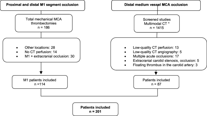
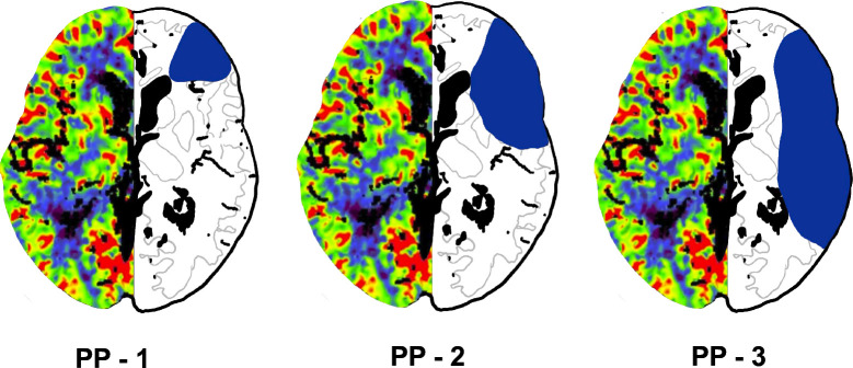
Methods: This retrospective study included 201 patients with acute ischaemic stroke, categorised into two groups: those with M1 segment occlusions (n=114) and those with distal medium vessel occlusions (n=87). We analysed multimodal stroke CT imaging and clinical data, focusing on the occlusion site, hypoperfusion extent and basal ganglia involvement. Patients with tandem stenosis or multiple acute occlusions were excluded. Perfusion patterns were categorised into three types (PP-1, PP-2 and PP-3) based on the extent of hypoperfusion. Statistical analysis explored associations between the occlusion site, perfusion pattern and collateral status.
Results: Among the 201 patients (mean age 75±14 years, 86 men), PP-1 was observed in 36.8% of patients (74/201), PP-2 in 27.4% (55/201) and PP-3 in 35.8% (72/201). The distribution of PP varied significantly by occlusion site (p<0.0001). Distal medium vessel occlusions were associated with PP-1 in 78.4% of cases (58/74), while PP-3 was most prevalent in M1 occlusions (90.3%, 65/72). The contingency coefficient revealed that occlusion location had a stronger association with the perfusion pattern (c=0.556) than collateral type (c=0.245). However, 21.6% of M1 occlusions (16/74) showed a PP-1 pattern and 9.7% of distal medium vessel occlusions (7/72) exhibited PP-3. Basal ganglia infarction presence was a reliable indicator of M1 occlusion with a 94% likelihood.
Conclusions: Perfusion patterns can effectively differentiate between proximal and distal medium vessel MCA occlusions, aiding targeted assessment of CT angiography.

 求助内容:
求助内容: 应助结果提醒方式:


