Philipp Kriechling, Jethin Joshy, Stefan Klotz, Georg C Feuerriegel, Philipp Fürnstahl, Reto Sutter, Mazda Farshad, Karl Wieser
{"title":"成功和失败的肩袖关节镜修复术后 5 年随访的三维肌肉体积和三维脂肪比例。","authors":"Philipp Kriechling, Jethin Joshy, Stefan Klotz, Georg C Feuerriegel, Philipp Fürnstahl, Reto Sutter, Mazda Farshad, Karl Wieser","doi":"10.1177/03635465241299795","DOIUrl":null,"url":null,"abstract":"<p><strong>Background: </strong>The results of rotator cuff (RC) repair surgery can be influenced by the presence of muscle atrophy and fatty infiltration. Three-dimensional (3D) quantitative data regarding those degenerative muscle changes after successful or failed RC repair are rare in the current literature.</p><p><strong>Hypothesis/purpose: </strong>The purpose of this study was to analyze muscle volume and fatty infiltration of the supraspinatus muscle after successful and failed arthroscopic RC tendon repair, with a minimum follow-up of 5 years. It was hypothesized that RC muscle volume and fatty infiltration would improve after successful repair and only to a limited extent after failed repair.</p><p><strong>Study design: </strong>Cohort study; Level of evidence 2.</p><p><strong>Methods: </strong>A total of 115 patients (mean age, 59 ± 8 years; 33% women) with RC repair for full-thickness supraspinatus tendon tear were prospectively followed at 3 and 12 months. Of them, 18 patients with unsuccessful RC repairs were matched to 21 patients with successful repairs and reevaluated at a minimum follow-up of 60 months. All patients underwent quantitative 2-point Dixon magnetic resonance imaging at all time points to evaluate full 3D muscle volume and 3D fatty infiltration. The clinical examination included the full Constant-Murley score (CS) and subjective shoulder value.</p><p><strong>Results: </strong>The relative changes in supraspinatus muscle volume were statistically significant between the 2 groups over time (<i>P</i> < .01). Successful repairs showed a mean volume increase of 18% (<i>P</i> < .001) and 23% (<i>P</i> < .001) from preoperatively and the 3-month follow-up, respectively, and failed repairs were remodeled by 3% (<i>P</i> = .586) and 12% (<i>P</i> = .001), respectively. However, a direct comparison of the volumes revealed comparable results at the latest follow-up with 43 cm<sup>3</sup> (95% CI, 38-47 cm<sup>3</sup>) and 40 cm<sup>3</sup> (95% CI, 33-46 cm<sup>3</sup>) for successful and failed repairs (<i>P</i> = .494), respectively. The supraspinatus 3D fatty infiltration also showed lower fat content for the successful repair preoperatively (6.9% [95% CI, 4.7%-9.2%] vs 9.1% [95% CI, 7.2%-11.1%]; <i>P</i> < .01), at 3 months (7.9% [95% CI, 5.5%-10.4%] vs 12.8% [95% CI, 9.1%-16.5%]; <i>P</i> < .01), at 12 months (7.5% [95% CI, 4.8%-10.1%] vs 11.6% [95% CI, 9.4%-13.8%]; <i>P</i> < .01), and at 60 months (7.4% [95% CI, 4.7%-10.2%] vs 15.5% [95% CI, 11%-20%]; <i>P</i> < .01) postoperatively. Fatty infiltration remained unchanged between preoperatively and from 3-month follow-up in the successful group. However, it increased by 70% (<i>P</i> < .01) from preoperative and by 21% (<i>P</i> = .065) from 3-months follow-up in the failed group. The clinical outcome was similar for successful and failed repairs with an absolute CS of 81 ± 6 versus 72 ± 15 (<i>P</i> = .069) and a relative CS of 94% ± 7% versus 85% ± 17% (<i>P</i> = .078).</p><p><strong>Conclusion: </strong>Successful RC repair was associated with relevant improvement of supraspinatus muscle mass and an unchanged fatty infiltration at a midterm follow-up of 5 years. However, failed repairs achieved only mild improvement of supraspinatus muscle volume and showed deterioration of fatty infiltration.</p>","PeriodicalId":55528,"journal":{"name":"American Journal of Sports Medicine","volume":"53 3","pages":"571-582"},"PeriodicalIF":4.5000,"publicationDate":"2025-03-01","publicationTypes":"Journal Article","fieldsOfStudy":null,"isOpenAccess":false,"openAccessPdf":"https://www.ncbi.nlm.nih.gov/pmc/articles/PMC11874612/pdf/","citationCount":"0","resultStr":"{\"title\":\"3D Muscle Volume and 3D Fat Fraction After Successful and Failed Arthroscopic Rotator Cuff Repair at 5-Year Follow-up.\",\"authors\":\"Philipp Kriechling, Jethin Joshy, Stefan Klotz, Georg C Feuerriegel, Philipp Fürnstahl, Reto Sutter, Mazda Farshad, Karl Wieser\",\"doi\":\"10.1177/03635465241299795\",\"DOIUrl\":null,\"url\":null,\"abstract\":\"<p><strong>Background: </strong>The results of rotator cuff (RC) repair surgery can be influenced by the presence of muscle atrophy and fatty infiltration. Three-dimensional (3D) quantitative data regarding those degenerative muscle changes after successful or failed RC repair are rare in the current literature.</p><p><strong>Hypothesis/purpose: </strong>The purpose of this study was to analyze muscle volume and fatty infiltration of the supraspinatus muscle after successful and failed arthroscopic RC tendon repair, with a minimum follow-up of 5 years. It was hypothesized that RC muscle volume and fatty infiltration would improve after successful repair and only to a limited extent after failed repair.</p><p><strong>Study design: </strong>Cohort study; Level of evidence 2.</p><p><strong>Methods: </strong>A total of 115 patients (mean age, 59 ± 8 years; 33% women) with RC repair for full-thickness supraspinatus tendon tear were prospectively followed at 3 and 12 months. Of them, 18 patients with unsuccessful RC repairs were matched to 21 patients with successful repairs and reevaluated at a minimum follow-up of 60 months. All patients underwent quantitative 2-point Dixon magnetic resonance imaging at all time points to evaluate full 3D muscle volume and 3D fatty infiltration. The clinical examination included the full Constant-Murley score (CS) and subjective shoulder value.</p><p><strong>Results: </strong>The relative changes in supraspinatus muscle volume were statistically significant between the 2 groups over time (<i>P</i> < .01). Successful repairs showed a mean volume increase of 18% (<i>P</i> < .001) and 23% (<i>P</i> < .001) from preoperatively and the 3-month follow-up, respectively, and failed repairs were remodeled by 3% (<i>P</i> = .586) and 12% (<i>P</i> = .001), respectively. However, a direct comparison of the volumes revealed comparable results at the latest follow-up with 43 cm<sup>3</sup> (95% CI, 38-47 cm<sup>3</sup>) and 40 cm<sup>3</sup> (95% CI, 33-46 cm<sup>3</sup>) for successful and failed repairs (<i>P</i> = .494), respectively. The supraspinatus 3D fatty infiltration also showed lower fat content for the successful repair preoperatively (6.9% [95% CI, 4.7%-9.2%] vs 9.1% [95% CI, 7.2%-11.1%]; <i>P</i> < .01), at 3 months (7.9% [95% CI, 5.5%-10.4%] vs 12.8% [95% CI, 9.1%-16.5%]; <i>P</i> < .01), at 12 months (7.5% [95% CI, 4.8%-10.1%] vs 11.6% [95% CI, 9.4%-13.8%]; <i>P</i> < .01), and at 60 months (7.4% [95% CI, 4.7%-10.2%] vs 15.5% [95% CI, 11%-20%]; <i>P</i> < .01) postoperatively. Fatty infiltration remained unchanged between preoperatively and from 3-month follow-up in the successful group. However, it increased by 70% (<i>P</i> < .01) from preoperative and by 21% (<i>P</i> = .065) from 3-months follow-up in the failed group. The clinical outcome was similar for successful and failed repairs with an absolute CS of 81 ± 6 versus 72 ± 15 (<i>P</i> = .069) and a relative CS of 94% ± 7% versus 85% ± 17% (<i>P</i> = .078).</p><p><strong>Conclusion: </strong>Successful RC repair was associated with relevant improvement of supraspinatus muscle mass and an unchanged fatty infiltration at a midterm follow-up of 5 years. However, failed repairs achieved only mild improvement of supraspinatus muscle volume and showed deterioration of fatty infiltration.</p>\",\"PeriodicalId\":55528,\"journal\":{\"name\":\"American Journal of Sports Medicine\",\"volume\":\"53 3\",\"pages\":\"571-582\"},\"PeriodicalIF\":4.5000,\"publicationDate\":\"2025-03-01\",\"publicationTypes\":\"Journal Article\",\"fieldsOfStudy\":null,\"isOpenAccess\":false,\"openAccessPdf\":\"https://www.ncbi.nlm.nih.gov/pmc/articles/PMC11874612/pdf/\",\"citationCount\":\"0\",\"resultStr\":null,\"platform\":\"Semanticscholar\",\"paperid\":null,\"PeriodicalName\":\"American Journal of Sports Medicine\",\"FirstCategoryId\":\"3\",\"ListUrlMain\":\"https://doi.org/10.1177/03635465241299795\",\"RegionNum\":1,\"RegionCategory\":\"医学\",\"ArticlePicture\":[],\"TitleCN\":null,\"AbstractTextCN\":null,\"PMCID\":null,\"EPubDate\":\"\",\"PubModel\":\"\",\"JCR\":\"Q1\",\"JCRName\":\"ORTHOPEDICS\",\"Score\":null,\"Total\":0}","platform":"Semanticscholar","paperid":null,"PeriodicalName":"American Journal of Sports Medicine","FirstCategoryId":"3","ListUrlMain":"https://doi.org/10.1177/03635465241299795","RegionNum":1,"RegionCategory":"医学","ArticlePicture":[],"TitleCN":null,"AbstractTextCN":null,"PMCID":null,"EPubDate":"","PubModel":"","JCR":"Q1","JCRName":"ORTHOPEDICS","Score":null,"Total":0}
3D Muscle Volume and 3D Fat Fraction After Successful and Failed Arthroscopic Rotator Cuff Repair at 5-Year Follow-up.
Background: The results of rotator cuff (RC) repair surgery can be influenced by the presence of muscle atrophy and fatty infiltration. Three-dimensional (3D) quantitative data regarding those degenerative muscle changes after successful or failed RC repair are rare in the current literature.
Hypothesis/purpose: The purpose of this study was to analyze muscle volume and fatty infiltration of the supraspinatus muscle after successful and failed arthroscopic RC tendon repair, with a minimum follow-up of 5 years. It was hypothesized that RC muscle volume and fatty infiltration would improve after successful repair and only to a limited extent after failed repair.
Study design: Cohort study; Level of evidence 2.
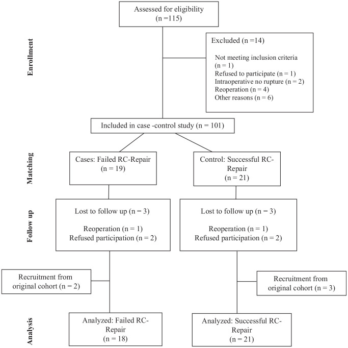
Methods: A total of 115 patients (mean age, 59 ± 8 years; 33% women) with RC repair for full-thickness supraspinatus tendon tear were prospectively followed at 3 and 12 months. Of them, 18 patients with unsuccessful RC repairs were matched to 21 patients with successful repairs and reevaluated at a minimum follow-up of 60 months. All patients underwent quantitative 2-point Dixon magnetic resonance imaging at all time points to evaluate full 3D muscle volume and 3D fatty infiltration. The clinical examination included the full Constant-Murley score (CS) and subjective shoulder value.
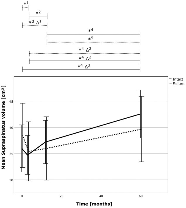
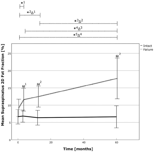
Results: The relative changes in supraspinatus muscle volume were statistically significant between the 2 groups over time (P < .01). Successful repairs showed a mean volume increase of 18% (P < .001) and 23% (P < .001) from preoperatively and the 3-month follow-up, respectively, and failed repairs were remodeled by 3% (P = .586) and 12% (P = .001), respectively. However, a direct comparison of the volumes revealed comparable results at the latest follow-up with 43 cm3 (95% CI, 38-47 cm3) and 40 cm3 (95% CI, 33-46 cm3) for successful and failed repairs (P = .494), respectively. The supraspinatus 3D fatty infiltration also showed lower fat content for the successful repair preoperatively (6.9% [95% CI, 4.7%-9.2%] vs 9.1% [95% CI, 7.2%-11.1%]; P < .01), at 3 months (7.9% [95% CI, 5.5%-10.4%] vs 12.8% [95% CI, 9.1%-16.5%]; P < .01), at 12 months (7.5% [95% CI, 4.8%-10.1%] vs 11.6% [95% CI, 9.4%-13.8%]; P < .01), and at 60 months (7.4% [95% CI, 4.7%-10.2%] vs 15.5% [95% CI, 11%-20%]; P < .01) postoperatively. Fatty infiltration remained unchanged between preoperatively and from 3-month follow-up in the successful group. However, it increased by 70% (P < .01) from preoperative and by 21% (P = .065) from 3-months follow-up in the failed group. The clinical outcome was similar for successful and failed repairs with an absolute CS of 81 ± 6 versus 72 ± 15 (P = .069) and a relative CS of 94% ± 7% versus 85% ± 17% (P = .078).
Conclusion: Successful RC repair was associated with relevant improvement of supraspinatus muscle mass and an unchanged fatty infiltration at a midterm follow-up of 5 years. However, failed repairs achieved only mild improvement of supraspinatus muscle volume and showed deterioration of fatty infiltration.
期刊介绍:
An invaluable resource for the orthopaedic sports medicine community, _The American Journal of Sports Medicine_ is a peer-reviewed scientific journal, first published in 1972. It is the official publication of the [American Orthopaedic Society for Sports Medicine (AOSSM)](http://www.sportsmed.org/)! The journal acts as an important forum for independent orthopaedic sports medicine research and education, allowing clinical practitioners the ability to make decisions based on sound scientific information.
This journal is a must-read for:
* Orthopaedic Surgeons and Specialists
* Sports Medicine Physicians
* Physiatrists
* Athletic Trainers
* Team Physicians
* And Physical Therapists
 求助内容:
求助内容: 应助结果提醒方式:


