Simona Ruxandra Volovăț, Diana-Ioana Boboc, Mădălina-Raluca Ostafe, Călin Gheorghe Buzea, Maricel Agop, Lăcrămioara Ochiuz, Dragoș Ioan Rusu, Decebal Vasincu, Monica Iuliana Ungureanu, Cristian Constantin Volovăț
{"title":"利用视觉变压器预测脑转移对磁共振成像引导阶段伽玛刀放射治疗的早期反应。","authors":"Simona Ruxandra Volovăț, Diana-Ioana Boboc, Mădălina-Raluca Ostafe, Călin Gheorghe Buzea, Maricel Agop, Lăcrămioara Ochiuz, Dragoș Ioan Rusu, Decebal Vasincu, Monica Iuliana Ungureanu, Cristian Constantin Volovăț","doi":"10.3390/tomography11020015","DOIUrl":null,"url":null,"abstract":"<p><strong>Background/objectives: </strong>This study explores the application of vision transformers to predict early responses to stereotactic radiosurgery in patients with brain metastases using minimally pre-processed magnetic resonance imaging scans. The objective is to assess the potential of vision transformers as a predictive tool for clinical decision-making, particularly in the context of imbalanced datasets.</p><p><strong>Methods: </strong>We analyzed magnetic resonance imaging scans from 19 brain metastases patients, focusing on axial fluid-attenuated inversion recovery and high-resolution contrast-enhanced T1-weighted sequences. Patients were categorized into responders (complete or partial response) and non-responders (stable or progressive disease).</p><p><strong>Results: </strong>Despite the imbalanced nature of the dataset, our results demonstrate that vision transformers can predict early treatment responses with an overall accuracy of 99%. The model exhibited high precision (99% for progression and 100% for regression) and recall (99% for progression and 100% for regression). The use of the attention mechanism in the vision transformers allowed the model to focus on relevant features in the magnetic resonance imaging images, ensuring an unbiased performance even with the imbalanced data. Confusion matrix analysis further confirmed the model's reliability, with minimal misclassifications. Additionally, the model achieved a perfect area under the receiver operator characteristic curve (AUC = 1.00), effectively distinguishing between responders and non-responders.</p><p><strong>Conclusions: </strong>These findings highlight the potential of vision transformers, aided by the attention mechanism, as a non-invasive, predictive tool for early response assessment in clinical oncology. The vision transformer (ViT) model employed in this study processes MRIs as sequences of patches, enabling the capture of localized tumor features critical for early response prediction. By leveraging patch-based feature learning, this approach enhances robustness, interpretability, and clinical applicability, addressing key challenges in tumor progression prediction following stereotactic radiosurgery (SRS). The model's robust performance, despite the dataset imbalance, underscores its ability to provide unbiased predictions. This approach could significantly enhance clinical decision-making and support personalized treatment strategies for brain metastases. Future research should validate these findings in larger, more diverse cohorts and explore the integration of additional data types to further optimize the model's clinical utility.</p>","PeriodicalId":51330,"journal":{"name":"Tomography","volume":"11 2","pages":""},"PeriodicalIF":2.2000,"publicationDate":"2025-02-07","publicationTypes":"Journal Article","fieldsOfStudy":null,"isOpenAccess":false,"openAccessPdf":"https://www.ncbi.nlm.nih.gov/pmc/articles/PMC11860310/pdf/","citationCount":"0","resultStr":"{\"title\":\"Utilizing Vision Transformers for Predicting Early Response of Brain Metastasis to Magnetic Resonance Imaging-Guided Stage Gamma Knife Radiosurgery Treatment.\",\"authors\":\"Simona Ruxandra Volovăț, Diana-Ioana Boboc, Mădălina-Raluca Ostafe, Călin Gheorghe Buzea, Maricel Agop, Lăcrămioara Ochiuz, Dragoș Ioan Rusu, Decebal Vasincu, Monica Iuliana Ungureanu, Cristian Constantin Volovăț\",\"doi\":\"10.3390/tomography11020015\",\"DOIUrl\":null,\"url\":null,\"abstract\":\"<p><strong>Background/objectives: </strong>This study explores the application of vision transformers to predict early responses to stereotactic radiosurgery in patients with brain metastases using minimally pre-processed magnetic resonance imaging scans. The objective is to assess the potential of vision transformers as a predictive tool for clinical decision-making, particularly in the context of imbalanced datasets.</p><p><strong>Methods: </strong>We analyzed magnetic resonance imaging scans from 19 brain metastases patients, focusing on axial fluid-attenuated inversion recovery and high-resolution contrast-enhanced T1-weighted sequences. Patients were categorized into responders (complete or partial response) and non-responders (stable or progressive disease).</p><p><strong>Results: </strong>Despite the imbalanced nature of the dataset, our results demonstrate that vision transformers can predict early treatment responses with an overall accuracy of 99%. The model exhibited high precision (99% for progression and 100% for regression) and recall (99% for progression and 100% for regression). The use of the attention mechanism in the vision transformers allowed the model to focus on relevant features in the magnetic resonance imaging images, ensuring an unbiased performance even with the imbalanced data. Confusion matrix analysis further confirmed the model's reliability, with minimal misclassifications. Additionally, the model achieved a perfect area under the receiver operator characteristic curve (AUC = 1.00), effectively distinguishing between responders and non-responders.</p><p><strong>Conclusions: </strong>These findings highlight the potential of vision transformers, aided by the attention mechanism, as a non-invasive, predictive tool for early response assessment in clinical oncology. The vision transformer (ViT) model employed in this study processes MRIs as sequences of patches, enabling the capture of localized tumor features critical for early response prediction. By leveraging patch-based feature learning, this approach enhances robustness, interpretability, and clinical applicability, addressing key challenges in tumor progression prediction following stereotactic radiosurgery (SRS). The model's robust performance, despite the dataset imbalance, underscores its ability to provide unbiased predictions. This approach could significantly enhance clinical decision-making and support personalized treatment strategies for brain metastases. Future research should validate these findings in larger, more diverse cohorts and explore the integration of additional data types to further optimize the model's clinical utility.</p>\",\"PeriodicalId\":51330,\"journal\":{\"name\":\"Tomography\",\"volume\":\"11 2\",\"pages\":\"\"},\"PeriodicalIF\":2.2000,\"publicationDate\":\"2025-02-07\",\"publicationTypes\":\"Journal Article\",\"fieldsOfStudy\":null,\"isOpenAccess\":false,\"openAccessPdf\":\"https://www.ncbi.nlm.nih.gov/pmc/articles/PMC11860310/pdf/\",\"citationCount\":\"0\",\"resultStr\":null,\"platform\":\"Semanticscholar\",\"paperid\":null,\"PeriodicalName\":\"Tomography\",\"FirstCategoryId\":\"3\",\"ListUrlMain\":\"https://doi.org/10.3390/tomography11020015\",\"RegionNum\":4,\"RegionCategory\":\"医学\",\"ArticlePicture\":[],\"TitleCN\":null,\"AbstractTextCN\":null,\"PMCID\":null,\"EPubDate\":\"\",\"PubModel\":\"\",\"JCR\":\"Q2\",\"JCRName\":\"RADIOLOGY, NUCLEAR MEDICINE & MEDICAL IMAGING\",\"Score\":null,\"Total\":0}","platform":"Semanticscholar","paperid":null,"PeriodicalName":"Tomography","FirstCategoryId":"3","ListUrlMain":"https://doi.org/10.3390/tomography11020015","RegionNum":4,"RegionCategory":"医学","ArticlePicture":[],"TitleCN":null,"AbstractTextCN":null,"PMCID":null,"EPubDate":"","PubModel":"","JCR":"Q2","JCRName":"RADIOLOGY, NUCLEAR MEDICINE & MEDICAL IMAGING","Score":null,"Total":0}
Utilizing Vision Transformers for Predicting Early Response of Brain Metastasis to Magnetic Resonance Imaging-Guided Stage Gamma Knife Radiosurgery Treatment.
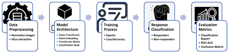
Background/objectives: This study explores the application of vision transformers to predict early responses to stereotactic radiosurgery in patients with brain metastases using minimally pre-processed magnetic resonance imaging scans. The objective is to assess the potential of vision transformers as a predictive tool for clinical decision-making, particularly in the context of imbalanced datasets.
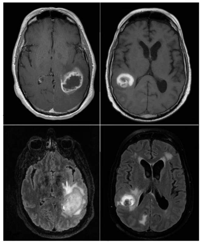
Methods: We analyzed magnetic resonance imaging scans from 19 brain metastases patients, focusing on axial fluid-attenuated inversion recovery and high-resolution contrast-enhanced T1-weighted sequences. Patients were categorized into responders (complete or partial response) and non-responders (stable or progressive disease).
Results: Despite the imbalanced nature of the dataset, our results demonstrate that vision transformers can predict early treatment responses with an overall accuracy of 99%. The model exhibited high precision (99% for progression and 100% for regression) and recall (99% for progression and 100% for regression). The use of the attention mechanism in the vision transformers allowed the model to focus on relevant features in the magnetic resonance imaging images, ensuring an unbiased performance even with the imbalanced data. Confusion matrix analysis further confirmed the model's reliability, with minimal misclassifications. Additionally, the model achieved a perfect area under the receiver operator characteristic curve (AUC = 1.00), effectively distinguishing between responders and non-responders.
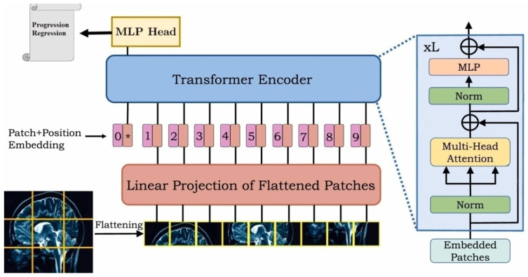
Conclusions: These findings highlight the potential of vision transformers, aided by the attention mechanism, as a non-invasive, predictive tool for early response assessment in clinical oncology. The vision transformer (ViT) model employed in this study processes MRIs as sequences of patches, enabling the capture of localized tumor features critical for early response prediction. By leveraging patch-based feature learning, this approach enhances robustness, interpretability, and clinical applicability, addressing key challenges in tumor progression prediction following stereotactic radiosurgery (SRS). The model's robust performance, despite the dataset imbalance, underscores its ability to provide unbiased predictions. This approach could significantly enhance clinical decision-making and support personalized treatment strategies for brain metastases. Future research should validate these findings in larger, more diverse cohorts and explore the integration of additional data types to further optimize the model's clinical utility.
TomographyMedicine-Radiology, Nuclear Medicine and Imaging
CiteScore
2.70
自引率
10.50%
发文量
222
期刊介绍:
TomographyTM publishes basic (technical and pre-clinical) and clinical scientific articles which involve the advancement of imaging technologies. Tomography encompasses studies that use single or multiple imaging modalities including for example CT, US, PET, SPECT, MR and hyperpolarization technologies, as well as optical modalities (i.e. bioluminescence, photoacoustic, endomicroscopy, fiber optic imaging and optical computed tomography) in basic sciences, engineering, preclinical and clinical medicine.
Tomography also welcomes studies involving exploration and refinement of contrast mechanisms and image-derived metrics within and across modalities toward the development of novel imaging probes for image-based feedback and intervention. The use of imaging in biology and medicine provides unparalleled opportunities to noninvasively interrogate tissues to obtain real-time dynamic and quantitative information required for diagnosis and response to interventions and to follow evolving pathological conditions. As multi-modal studies and the complexities of imaging technologies themselves are ever increasing to provide advanced information to scientists and clinicians.
Tomography provides a unique publication venue allowing investigators the opportunity to more precisely communicate integrated findings related to the diverse and heterogeneous features associated with underlying anatomical, physiological, functional, metabolic and molecular genetic activities of normal and diseased tissue. Thus Tomography publishes peer-reviewed articles which involve the broad use of imaging of any tissue and disease type including both preclinical and clinical investigations. In addition, hardware/software along with chemical and molecular probe advances are welcome as they are deemed to significantly contribute towards the long-term goal of improving the overall impact of imaging on scientific and clinical discovery.

 求助内容:
求助内容: 应助结果提醒方式:


