Wolfgang Wirth, Simon Herger, Susanne Maschek, Anna Wisser, Oliver Bieri, Felix Eckstein, Annegret Mündermann
{"title":"全自动软骨横向松弛时间(T2)和厚度定量DESS磁共振成像的临床验证。","authors":"Wolfgang Wirth, Simon Herger, Susanne Maschek, Anna Wisser, Oliver Bieri, Felix Eckstein, Annegret Mündermann","doi":"10.1007/s10334-025-01227-5","DOIUrl":null,"url":null,"abstract":"<p><strong>Objective: </strong>To clinically validate a fully automated cartilage segmentation technique from quantitative double-echo steady-state (qDESS) MRI supporting simultaneous estimation of cartilage T2 and morphology. Here, we test whether laminar (superficial and deep layer) T2 results from convolutional neural network (CNN) segmentations are consistent with those from manual expert segmentations.</p><p><strong>Materials and methods: </strong>The 3D qDESS sequence was acquired using 3 T MRI (resolution: 0.3125 × 0.3125x1.5 mm) in both knees of 37 subjects with unilateral anterior cruciate ligament (ACL) injury and 48 uninjured controls. Automated femorotibial cartilage (FTJ) segmentation was based on a 2D U-Net. Laminar T2 and cartilage thickness across the FTJ) were compared between ACL-injured and contralateral knees, and between ACL-injured and control knees. Effect sizes of these differences were measured using non-parametric Cohen's d (d<sub>n-p</sub>).</p><p><strong>Result: </strong>Significant differences were observed only in deep T2, with longer T2 in ACL-injured knees than in contralateral and healthy control knees in most of the comparisons and with similar effect sizes for automated and manual segmentations (range d<sub>n-p</sub> automated/manual: 0.58-1.04/0.58-0.74). No significant differences were observed in superficial T2 or cartilage thickness.</p><p><strong>Discussion: </strong>Fully-automated, CNN-based analysis showed similar sensitivity to differences in laminar cartilage T2 as manual segmentation, allowing automated qDESS-analyses to be applied to larger datasets.</p>","PeriodicalId":18067,"journal":{"name":"Magnetic Resonance Materials in Physics, Biology and Medicine","volume":" ","pages":"285-297"},"PeriodicalIF":2.5000,"publicationDate":"2025-04-01","publicationTypes":"Journal Article","fieldsOfStudy":null,"isOpenAccess":false,"openAccessPdf":"https://www.ncbi.nlm.nih.gov/pmc/articles/PMC11914229/pdf/","citationCount":"0","resultStr":"{\"title\":\"Clinical validation of fully automated cartilage transverse relaxation time (T2) and thickness analysis using quantitative DESS magnetic resonance imaging.\",\"authors\":\"Wolfgang Wirth, Simon Herger, Susanne Maschek, Anna Wisser, Oliver Bieri, Felix Eckstein, Annegret Mündermann\",\"doi\":\"10.1007/s10334-025-01227-5\",\"DOIUrl\":null,\"url\":null,\"abstract\":\"<p><strong>Objective: </strong>To clinically validate a fully automated cartilage segmentation technique from quantitative double-echo steady-state (qDESS) MRI supporting simultaneous estimation of cartilage T2 and morphology. Here, we test whether laminar (superficial and deep layer) T2 results from convolutional neural network (CNN) segmentations are consistent with those from manual expert segmentations.</p><p><strong>Materials and methods: </strong>The 3D qDESS sequence was acquired using 3 T MRI (resolution: 0.3125 × 0.3125x1.5 mm) in both knees of 37 subjects with unilateral anterior cruciate ligament (ACL) injury and 48 uninjured controls. Automated femorotibial cartilage (FTJ) segmentation was based on a 2D U-Net. Laminar T2 and cartilage thickness across the FTJ) were compared between ACL-injured and contralateral knees, and between ACL-injured and control knees. Effect sizes of these differences were measured using non-parametric Cohen's d (d<sub>n-p</sub>).</p><p><strong>Result: </strong>Significant differences were observed only in deep T2, with longer T2 in ACL-injured knees than in contralateral and healthy control knees in most of the comparisons and with similar effect sizes for automated and manual segmentations (range d<sub>n-p</sub> automated/manual: 0.58-1.04/0.58-0.74). No significant differences were observed in superficial T2 or cartilage thickness.</p><p><strong>Discussion: </strong>Fully-automated, CNN-based analysis showed similar sensitivity to differences in laminar cartilage T2 as manual segmentation, allowing automated qDESS-analyses to be applied to larger datasets.</p>\",\"PeriodicalId\":18067,\"journal\":{\"name\":\"Magnetic Resonance Materials in Physics, Biology and Medicine\",\"volume\":\" \",\"pages\":\"285-297\"},\"PeriodicalIF\":2.5000,\"publicationDate\":\"2025-04-01\",\"publicationTypes\":\"Journal Article\",\"fieldsOfStudy\":null,\"isOpenAccess\":false,\"openAccessPdf\":\"https://www.ncbi.nlm.nih.gov/pmc/articles/PMC11914229/pdf/\",\"citationCount\":\"0\",\"resultStr\":null,\"platform\":\"Semanticscholar\",\"paperid\":null,\"PeriodicalName\":\"Magnetic Resonance Materials in Physics, Biology and Medicine\",\"FirstCategoryId\":\"3\",\"ListUrlMain\":\"https://doi.org/10.1007/s10334-025-01227-5\",\"RegionNum\":4,\"RegionCategory\":\"医学\",\"ArticlePicture\":[],\"TitleCN\":null,\"AbstractTextCN\":null,\"PMCID\":null,\"EPubDate\":\"2025/2/24 0:00:00\",\"PubModel\":\"Epub\",\"JCR\":\"Q3\",\"JCRName\":\"RADIOLOGY, NUCLEAR MEDICINE & MEDICAL IMAGING\",\"Score\":null,\"Total\":0}","platform":"Semanticscholar","paperid":null,"PeriodicalName":"Magnetic Resonance Materials in Physics, Biology and Medicine","FirstCategoryId":"3","ListUrlMain":"https://doi.org/10.1007/s10334-025-01227-5","RegionNum":4,"RegionCategory":"医学","ArticlePicture":[],"TitleCN":null,"AbstractTextCN":null,"PMCID":null,"EPubDate":"2025/2/24 0:00:00","PubModel":"Epub","JCR":"Q3","JCRName":"RADIOLOGY, NUCLEAR MEDICINE & MEDICAL IMAGING","Score":null,"Total":0}
引用次数: 0
摘要
目的:临床验证定量双回声稳态(qDESS) MRI的全自动软骨分割技术,支持同时估计软骨T2和形态。在这里,我们测试了卷积神经网络(CNN)分割的层流(浅层和深层)T2结果与人工专家分割的结果是否一致。材料与方法:采用3t MRI(分辨率:0.3125 × 0.3125 × 1.5 mm)对37例单侧前交叉韧带(ACL)损伤的双膝和48例未损伤的对照组进行三维qDESS序列采集。自动股胫软骨(FTJ)分割基于二维U-Net。比较acl损伤膝关节与对侧膝关节、acl损伤膝关节与对照膝关节间的层间T2和FTJ软骨厚度。使用非参数Cohen's d (dn-p)测量这些差异的效应量。结果:在大多数比较中,只有深层T2存在显著差异,acl损伤膝关节的T2比对侧和健康对照膝关节的T2更长,自动和手动分割的效应大小相似(范围dn-p自动/手动:0.58-1.04/0.58-0.74)。两组在T2表浅及软骨厚度上均无明显差异。讨论:全自动的、基于cnn的分析显示出与人工分割相似的对板层软骨T2差异的敏感性,允许自动qdess分析应用于更大的数据集。
Clinical validation of fully automated cartilage transverse relaxation time (T2) and thickness analysis using quantitative DESS magnetic resonance imaging.
Objective: To clinically validate a fully automated cartilage segmentation technique from quantitative double-echo steady-state (qDESS) MRI supporting simultaneous estimation of cartilage T2 and morphology. Here, we test whether laminar (superficial and deep layer) T2 results from convolutional neural network (CNN) segmentations are consistent with those from manual expert segmentations.
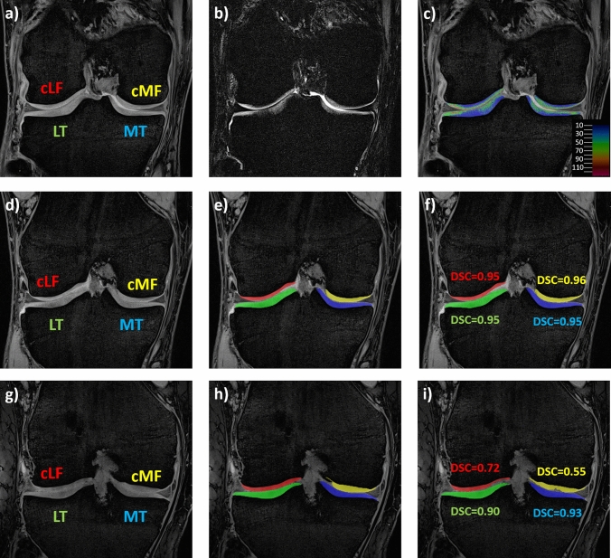
Materials and methods: The 3D qDESS sequence was acquired using 3 T MRI (resolution: 0.3125 × 0.3125x1.5 mm) in both knees of 37 subjects with unilateral anterior cruciate ligament (ACL) injury and 48 uninjured controls. Automated femorotibial cartilage (FTJ) segmentation was based on a 2D U-Net. Laminar T2 and cartilage thickness across the FTJ) were compared between ACL-injured and contralateral knees, and between ACL-injured and control knees. Effect sizes of these differences were measured using non-parametric Cohen's d (dn-p).
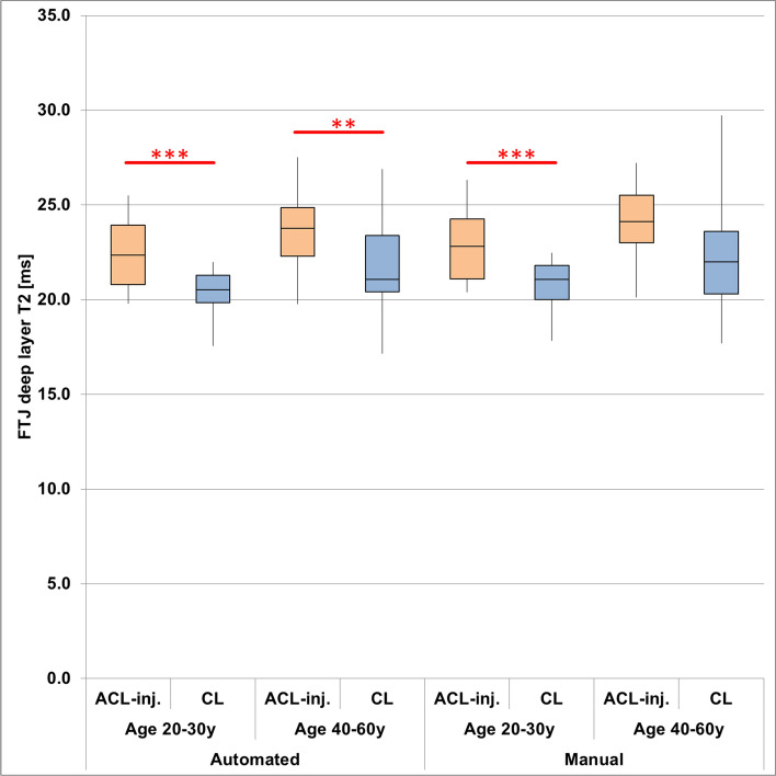
Result: Significant differences were observed only in deep T2, with longer T2 in ACL-injured knees than in contralateral and healthy control knees in most of the comparisons and with similar effect sizes for automated and manual segmentations (range dn-p automated/manual: 0.58-1.04/0.58-0.74). No significant differences were observed in superficial T2 or cartilage thickness.
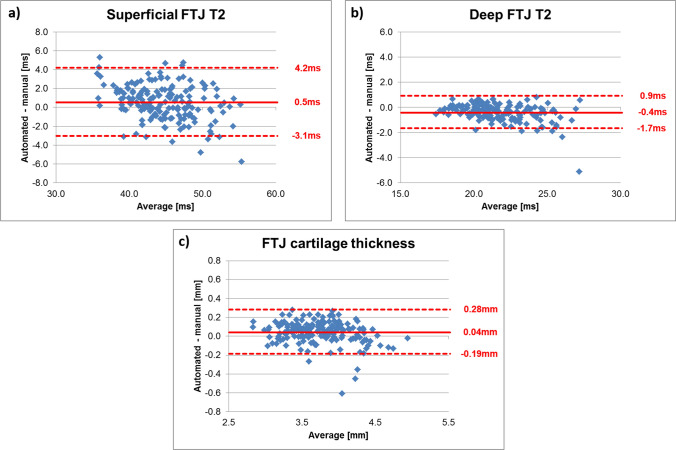
Discussion: Fully-automated, CNN-based analysis showed similar sensitivity to differences in laminar cartilage T2 as manual segmentation, allowing automated qDESS-analyses to be applied to larger datasets.
期刊介绍:
MAGMA is a multidisciplinary international journal devoted to the publication of articles on all aspects of magnetic resonance techniques and their applications in medicine and biology. MAGMA currently publishes research papers, reviews, letters to the editor, and commentaries, six times a year. The subject areas covered by MAGMA include:
advances in materials, hardware and software in magnetic resonance technology,
new developments and results in research and practical applications of magnetic resonance imaging and spectroscopy related to biology and medicine,
study of animal models and intact cells using magnetic resonance,
reports of clinical trials on humans and clinical validation of magnetic resonance protocols.

 求助内容:
求助内容: 应助结果提醒方式:


