Alison Maclean, Laura Tipple, Emily Newton, Dharani K Hapangama
{"title":"b子宫腺肌症患者异位和异位子宫内膜激素受体谱:系统综述。","authors":"Alison Maclean, Laura Tipple, Emily Newton, Dharani K Hapangama","doi":"10.1093/hropen/hoaf002","DOIUrl":null,"url":null,"abstract":"<p><strong>Study question: </strong>What is the hormone receptor profile of adenomyosis lesions in comparison to correctly located endometrium?</p><p><strong>Summary answer: </strong>Adenomyosis lesions exhibit increased oestrogen receptor (ER) expression compared to the eutopic endometrium; there are conflicting results regarding progesterone receptor (PR) expression and a lack of studies on androgen receptor (AR) expression.</p><p><strong>What is known already: </strong>Adenomyosis lesions express hormone receptors indicating an influence from ovarian steroid hormones. However, hormone treatments are often ineffective in controlling adenomyosis symptoms, which suggests alternate hormonal responses and, potentially, a distinct hormone receptor expression profile within adenomyosis lesions compared to the eutopic endometrium.</p><p><strong>Study design size duration: </strong>This systematic review with a thematic analysis retrieved studies from the PubMed, Ovid Medline, Embase, Scopus, and Cochrane Library databases, and searches were conducted from inception through to May 2024. Human studies were included and identified using a combination of exploded MeSH terms ('adenomyosis') and free-text search terms ('oestrogen receptor', 'progesterone receptor', 'androgen receptor', 'hormone receptor').</p><p><strong>Participants/materials setting methods: </strong>This review was reported in accordance with the PRISMA guidelines. All studies reporting original data concerning hormone receptors in adenomyosis lesions compared to eutopic endometrium in adenomyosis were included. Studies that did not report original data or provide a review of the field were excluded. Bias analysis was completed for each study using the Newcastle-Ottawa scoring system.</p><p><strong>Main results and the role of chance: </strong>There were 1905 studies identified, which were screened to include 12 studies that met the eligibility criteria, including 11 proteomic studies and one transcriptional study, with a total of 555 individual participants. ER expression was consistently increased in adenomyosis lesions compared to the eutopic endometrium, specifically in the secretory phase. When endometrial subregion was considered, this difference was specific to the endometrial functionalis only. When different isoforms were considered, this increase in ER expression was specific to ERα rather than ERβ. There were conflicting results on PR expression, with most studies showing no significant difference or reduced levels in adenomyosis lesions compared to the eutopic endometrium. There is a paucity of data on AR expression in adenomyosis lesions, with only one study of small sample size included.</p><p><strong>Limitations reasons for caution: </strong>A high risk of bias arose from studies grouping endometrial samples across different menstrual cycle phases for analysis. The coexistence of gynecological conditions like endometriosis may also confound the hormone receptor profile of the eutopic endometrium. Most studies employing immunostaining did not comment on region-specific differences in the endometrium. Given the well-documented cyclical variations in hormone receptor expression within the endometrium, the need for more attention to region-specific differences represents a notable limitation in the current body of literature.</p><p><strong>Wider implications of the findings: </strong>The systematic review highlights oestrogen dominance through elevated ERα levels in adenomyosis lesions, which agrees with the literature suggesting local hyper-oestrogenism in adenomyosis lesions. Heterogeneity in menstrual cycle timing and lack of endometrial region specificity prevent conclusions on progesterone resistance within adenomyosis lesions in this study. Future investigations should minimize the bias through well-defined cohorts, leading to robust exploration of hormone receptor profiles in adenomyosis lesions to identify therapeutic targets and deepen our understanding of adenomyosis pathogenesis.</p><p><strong>Study funding/competing interests: </strong>This work was supported by Wellbeing of Women Research Project grants RG1073 and RG2137 (D.K.H.), a Wellbeing of Women Entry-Level Scholarship ELS706 and a Medical Research Council grant MR/V007238/1 (A.M. and D.K.H.), as well as the University of Liverpool (L.T.). There are no conflicts of interest.</p><p><strong>Hropen-24-0294r2: </strong>The review protocol was published in the PROSPERO Register of Systematic Reviews on 27 September 2023, registration number CRD4202346.</p>","PeriodicalId":73264,"journal":{"name":"Human reproduction open","volume":"2025 1","pages":"hoaf002"},"PeriodicalIF":11.1000,"publicationDate":"2025-01-20","publicationTypes":"Journal Article","fieldsOfStudy":null,"isOpenAccess":false,"openAccessPdf":"https://www.ncbi.nlm.nih.gov/pmc/articles/PMC11810641/pdf/","citationCount":"0","resultStr":"{\"title\":\"Hormone receptor profile of ectopic and eutopic endometrium in adenomyosis: a systematic review.\",\"authors\":\"Alison Maclean, Laura Tipple, Emily Newton, Dharani K Hapangama\",\"doi\":\"10.1093/hropen/hoaf002\",\"DOIUrl\":null,\"url\":null,\"abstract\":\"<p><strong>Study question: </strong>What is the hormone receptor profile of adenomyosis lesions in comparison to correctly located endometrium?</p><p><strong>Summary answer: </strong>Adenomyosis lesions exhibit increased oestrogen receptor (ER) expression compared to the eutopic endometrium; there are conflicting results regarding progesterone receptor (PR) expression and a lack of studies on androgen receptor (AR) expression.</p><p><strong>What is known already: </strong>Adenomyosis lesions express hormone receptors indicating an influence from ovarian steroid hormones. However, hormone treatments are often ineffective in controlling adenomyosis symptoms, which suggests alternate hormonal responses and, potentially, a distinct hormone receptor expression profile within adenomyosis lesions compared to the eutopic endometrium.</p><p><strong>Study design size duration: </strong>This systematic review with a thematic analysis retrieved studies from the PubMed, Ovid Medline, Embase, Scopus, and Cochrane Library databases, and searches were conducted from inception through to May 2024. Human studies were included and identified using a combination of exploded MeSH terms ('adenomyosis') and free-text search terms ('oestrogen receptor', 'progesterone receptor', 'androgen receptor', 'hormone receptor').</p><p><strong>Participants/materials setting methods: </strong>This review was reported in accordance with the PRISMA guidelines. All studies reporting original data concerning hormone receptors in adenomyosis lesions compared to eutopic endometrium in adenomyosis were included. Studies that did not report original data or provide a review of the field were excluded. Bias analysis was completed for each study using the Newcastle-Ottawa scoring system.</p><p><strong>Main results and the role of chance: </strong>There were 1905 studies identified, which were screened to include 12 studies that met the eligibility criteria, including 11 proteomic studies and one transcriptional study, with a total of 555 individual participants. ER expression was consistently increased in adenomyosis lesions compared to the eutopic endometrium, specifically in the secretory phase. When endometrial subregion was considered, this difference was specific to the endometrial functionalis only. When different isoforms were considered, this increase in ER expression was specific to ERα rather than ERβ. There were conflicting results on PR expression, with most studies showing no significant difference or reduced levels in adenomyosis lesions compared to the eutopic endometrium. There is a paucity of data on AR expression in adenomyosis lesions, with only one study of small sample size included.</p><p><strong>Limitations reasons for caution: </strong>A high risk of bias arose from studies grouping endometrial samples across different menstrual cycle phases for analysis. The coexistence of gynecological conditions like endometriosis may also confound the hormone receptor profile of the eutopic endometrium. Most studies employing immunostaining did not comment on region-specific differences in the endometrium. Given the well-documented cyclical variations in hormone receptor expression within the endometrium, the need for more attention to region-specific differences represents a notable limitation in the current body of literature.</p><p><strong>Wider implications of the findings: </strong>The systematic review highlights oestrogen dominance through elevated ERα levels in adenomyosis lesions, which agrees with the literature suggesting local hyper-oestrogenism in adenomyosis lesions. Heterogeneity in menstrual cycle timing and lack of endometrial region specificity prevent conclusions on progesterone resistance within adenomyosis lesions in this study. Future investigations should minimize the bias through well-defined cohorts, leading to robust exploration of hormone receptor profiles in adenomyosis lesions to identify therapeutic targets and deepen our understanding of adenomyosis pathogenesis.</p><p><strong>Study funding/competing interests: </strong>This work was supported by Wellbeing of Women Research Project grants RG1073 and RG2137 (D.K.H.), a Wellbeing of Women Entry-Level Scholarship ELS706 and a Medical Research Council grant MR/V007238/1 (A.M. and D.K.H.), as well as the University of Liverpool (L.T.). There are no conflicts of interest.</p><p><strong>Hropen-24-0294r2: </strong>The review protocol was published in the PROSPERO Register of Systematic Reviews on 27 September 2023, registration number CRD4202346.</p>\",\"PeriodicalId\":73264,\"journal\":{\"name\":\"Human reproduction open\",\"volume\":\"2025 1\",\"pages\":\"hoaf002\"},\"PeriodicalIF\":11.1000,\"publicationDate\":\"2025-01-20\",\"publicationTypes\":\"Journal Article\",\"fieldsOfStudy\":null,\"isOpenAccess\":false,\"openAccessPdf\":\"https://www.ncbi.nlm.nih.gov/pmc/articles/PMC11810641/pdf/\",\"citationCount\":\"0\",\"resultStr\":null,\"platform\":\"Semanticscholar\",\"paperid\":null,\"PeriodicalName\":\"Human reproduction open\",\"FirstCategoryId\":\"1085\",\"ListUrlMain\":\"https://doi.org/10.1093/hropen/hoaf002\",\"RegionNum\":0,\"RegionCategory\":null,\"ArticlePicture\":[],\"TitleCN\":null,\"AbstractTextCN\":null,\"PMCID\":null,\"EPubDate\":\"2025/1/1 0:00:00\",\"PubModel\":\"eCollection\",\"JCR\":\"Q1\",\"JCRName\":\"OBSTETRICS & GYNECOLOGY\",\"Score\":null,\"Total\":0}","platform":"Semanticscholar","paperid":null,"PeriodicalName":"Human reproduction open","FirstCategoryId":"1085","ListUrlMain":"https://doi.org/10.1093/hropen/hoaf002","RegionNum":0,"RegionCategory":null,"ArticlePicture":[],"TitleCN":null,"AbstractTextCN":null,"PMCID":null,"EPubDate":"2025/1/1 0:00:00","PubModel":"eCollection","JCR":"Q1","JCRName":"OBSTETRICS & GYNECOLOGY","Score":null,"Total":0}
Hormone receptor profile of ectopic and eutopic endometrium in adenomyosis: a systematic review.
Study question: What is the hormone receptor profile of adenomyosis lesions in comparison to correctly located endometrium?
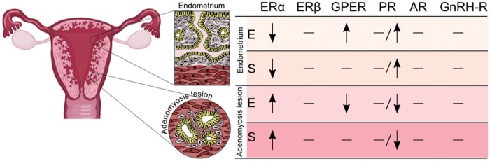
Summary answer: Adenomyosis lesions exhibit increased oestrogen receptor (ER) expression compared to the eutopic endometrium; there are conflicting results regarding progesterone receptor (PR) expression and a lack of studies on androgen receptor (AR) expression.
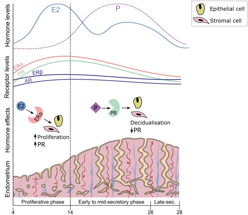
What is known already: Adenomyosis lesions express hormone receptors indicating an influence from ovarian steroid hormones. However, hormone treatments are often ineffective in controlling adenomyosis symptoms, which suggests alternate hormonal responses and, potentially, a distinct hormone receptor expression profile within adenomyosis lesions compared to the eutopic endometrium.
Study design size duration: This systematic review with a thematic analysis retrieved studies from the PubMed, Ovid Medline, Embase, Scopus, and Cochrane Library databases, and searches were conducted from inception through to May 2024. Human studies were included and identified using a combination of exploded MeSH terms ('adenomyosis') and free-text search terms ('oestrogen receptor', 'progesterone receptor', 'androgen receptor', 'hormone receptor').
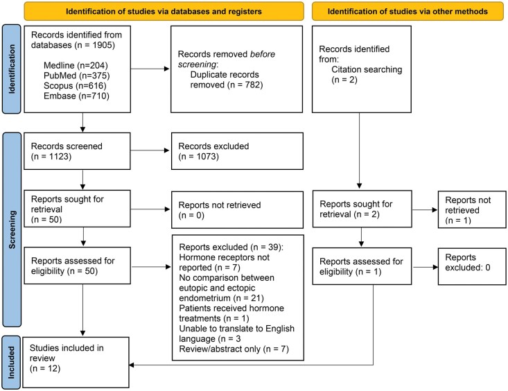
Participants/materials setting methods: This review was reported in accordance with the PRISMA guidelines. All studies reporting original data concerning hormone receptors in adenomyosis lesions compared to eutopic endometrium in adenomyosis were included. Studies that did not report original data or provide a review of the field were excluded. Bias analysis was completed for each study using the Newcastle-Ottawa scoring system.
Main results and the role of chance: There were 1905 studies identified, which were screened to include 12 studies that met the eligibility criteria, including 11 proteomic studies and one transcriptional study, with a total of 555 individual participants. ER expression was consistently increased in adenomyosis lesions compared to the eutopic endometrium, specifically in the secretory phase. When endometrial subregion was considered, this difference was specific to the endometrial functionalis only. When different isoforms were considered, this increase in ER expression was specific to ERα rather than ERβ. There were conflicting results on PR expression, with most studies showing no significant difference or reduced levels in adenomyosis lesions compared to the eutopic endometrium. There is a paucity of data on AR expression in adenomyosis lesions, with only one study of small sample size included.
Limitations reasons for caution: A high risk of bias arose from studies grouping endometrial samples across different menstrual cycle phases for analysis. The coexistence of gynecological conditions like endometriosis may also confound the hormone receptor profile of the eutopic endometrium. Most studies employing immunostaining did not comment on region-specific differences in the endometrium. Given the well-documented cyclical variations in hormone receptor expression within the endometrium, the need for more attention to region-specific differences represents a notable limitation in the current body of literature.
Wider implications of the findings: The systematic review highlights oestrogen dominance through elevated ERα levels in adenomyosis lesions, which agrees with the literature suggesting local hyper-oestrogenism in adenomyosis lesions. Heterogeneity in menstrual cycle timing and lack of endometrial region specificity prevent conclusions on progesterone resistance within adenomyosis lesions in this study. Future investigations should minimize the bias through well-defined cohorts, leading to robust exploration of hormone receptor profiles in adenomyosis lesions to identify therapeutic targets and deepen our understanding of adenomyosis pathogenesis.
Study funding/competing interests: This work was supported by Wellbeing of Women Research Project grants RG1073 and RG2137 (D.K.H.), a Wellbeing of Women Entry-Level Scholarship ELS706 and a Medical Research Council grant MR/V007238/1 (A.M. and D.K.H.), as well as the University of Liverpool (L.T.). There are no conflicts of interest.
Hropen-24-0294r2: The review protocol was published in the PROSPERO Register of Systematic Reviews on 27 September 2023, registration number CRD4202346.

 求助内容:
求助内容: 应助结果提醒方式:


