Sergiy Yakovlev*, David A. Nyenhuis, Nico Tjandra, Dudley K. Strickland and Leonid Medved*,
{"title":"纤维蛋白与n -钙粘蛋白相互作用关键氨基酸残基的鉴定","authors":"Sergiy Yakovlev*, David A. Nyenhuis, Nico Tjandra, Dudley K. Strickland and Leonid Medved*, ","doi":"10.1021/acs.biochem.4c0051010.1021/acs.biochem.4c00510","DOIUrl":null,"url":null,"abstract":"<p >We recently identified N-cadherin as a novel receptor for fibrin and localized complementary binding sites within the fibrin βN-domains and the third and fifth extracellular domains (EC3 and EC5) of N-cadherin. We also hypothesized that the His16 and Arg17 residues of the βN-domains and the (Asp/Glu)-X-(Asp/Glu) motifs present in the EC3 and EC5 domains may play roles in the interaction between fibrin and N-cadherin. The primary objectives of this study were to test these hypotheses and to further clarify the structural basis for this interaction. To test our hypotheses, we first mutated His16 and Arg17 in the recombinant (β15–66)<sub>2</sub> fragment, which mimics the dimeric arrangement of the βN-domains in fibrin, using site-directed mutagenesis. The results revealed that the mutations of both His16 and Arg17 are critical for the interaction. Next, we mutated Asp/Glu residues in the three (Asp/Glu)-X-(Asp/Glu) motifs, M1 (Asp-Phe-Glu), M2 (Glu-Ala-Glu), and M3 (Asp-Tyr-Asp), of the fibrin-binding N-cad(3–5) fragment of N-cadherin. The results showed that Asp292 and Glu294 of M1, and Asp468 and Asp470 of M3, are critical for the interaction. Our molecular modeling of the 3D structure of the EC3-EC4-EC5 domains revealed that these residues are located at the interfaces of EC3-EC4 and EC4-EC5 and that some may also be involved in calcium binding. In conclusion, our study identified amino acid residues in the fibrin βN-domains and the EC3 and EC5 domains of N-cadherin that are critical for the interaction of fibrin with N-cadherin and localized the fibrin-binding residues in the 3D structure of N-cadherin.</p>","PeriodicalId":28,"journal":{"name":"Biochemistry Biochemistry","volume":"64 1","pages":"83–91 83–91"},"PeriodicalIF":3.0000,"publicationDate":"2024-12-13","publicationTypes":"Journal Article","fieldsOfStudy":null,"isOpenAccess":false,"openAccessPdf":"","citationCount":"0","resultStr":"{\"title\":\"Identification of Amino Acid Residues Critical for the Interaction of Fibrin with N-Cadherin\",\"authors\":\"Sergiy Yakovlev*, David A. Nyenhuis, Nico Tjandra, Dudley K. Strickland and Leonid Medved*, \",\"doi\":\"10.1021/acs.biochem.4c0051010.1021/acs.biochem.4c00510\",\"DOIUrl\":null,\"url\":null,\"abstract\":\"<p >We recently identified N-cadherin as a novel receptor for fibrin and localized complementary binding sites within the fibrin βN-domains and the third and fifth extracellular domains (EC3 and EC5) of N-cadherin. We also hypothesized that the His16 and Arg17 residues of the βN-domains and the (Asp/Glu)-X-(Asp/Glu) motifs present in the EC3 and EC5 domains may play roles in the interaction between fibrin and N-cadherin. The primary objectives of this study were to test these hypotheses and to further clarify the structural basis for this interaction. To test our hypotheses, we first mutated His16 and Arg17 in the recombinant (β15–66)<sub>2</sub> fragment, which mimics the dimeric arrangement of the βN-domains in fibrin, using site-directed mutagenesis. The results revealed that the mutations of both His16 and Arg17 are critical for the interaction. Next, we mutated Asp/Glu residues in the three (Asp/Glu)-X-(Asp/Glu) motifs, M1 (Asp-Phe-Glu), M2 (Glu-Ala-Glu), and M3 (Asp-Tyr-Asp), of the fibrin-binding N-cad(3–5) fragment of N-cadherin. The results showed that Asp292 and Glu294 of M1, and Asp468 and Asp470 of M3, are critical for the interaction. Our molecular modeling of the 3D structure of the EC3-EC4-EC5 domains revealed that these residues are located at the interfaces of EC3-EC4 and EC4-EC5 and that some may also be involved in calcium binding. In conclusion, our study identified amino acid residues in the fibrin βN-domains and the EC3 and EC5 domains of N-cadherin that are critical for the interaction of fibrin with N-cadherin and localized the fibrin-binding residues in the 3D structure of N-cadherin.</p>\",\"PeriodicalId\":28,\"journal\":{\"name\":\"Biochemistry Biochemistry\",\"volume\":\"64 1\",\"pages\":\"83–91 83–91\"},\"PeriodicalIF\":3.0000,\"publicationDate\":\"2024-12-13\",\"publicationTypes\":\"Journal Article\",\"fieldsOfStudy\":null,\"isOpenAccess\":false,\"openAccessPdf\":\"\",\"citationCount\":\"0\",\"resultStr\":null,\"platform\":\"Semanticscholar\",\"paperid\":null,\"PeriodicalName\":\"Biochemistry Biochemistry\",\"FirstCategoryId\":\"1\",\"ListUrlMain\":\"https://pubs.acs.org/doi/10.1021/acs.biochem.4c00510\",\"RegionNum\":3,\"RegionCategory\":\"生物学\",\"ArticlePicture\":[],\"TitleCN\":null,\"AbstractTextCN\":null,\"PMCID\":null,\"EPubDate\":\"\",\"PubModel\":\"\",\"JCR\":\"Q3\",\"JCRName\":\"BIOCHEMISTRY & MOLECULAR BIOLOGY\",\"Score\":null,\"Total\":0}","platform":"Semanticscholar","paperid":null,"PeriodicalName":"Biochemistry Biochemistry","FirstCategoryId":"1","ListUrlMain":"https://pubs.acs.org/doi/10.1021/acs.biochem.4c00510","RegionNum":3,"RegionCategory":"生物学","ArticlePicture":[],"TitleCN":null,"AbstractTextCN":null,"PMCID":null,"EPubDate":"","PubModel":"","JCR":"Q3","JCRName":"BIOCHEMISTRY & MOLECULAR BIOLOGY","Score":null,"Total":0}
引用次数: 0
摘要
我们最近发现N-cadherin是一种新的纤维蛋白受体,并在纤维蛋白β n -结构域和N-cadherin的第三和第五胞外结构域(EC3和EC5)内定位了互补结合位点。我们还假设β n结构域的His16和Arg17残基以及EC3和EC5结构域的(Asp/Glu)- x -(Asp/Glu)基序可能在纤维蛋白和n -钙粘蛋白之间的相互作用中发挥作用。本研究的主要目的是检验这些假设,并进一步阐明这种相互作用的结构基础。为了验证我们的假设,我们首先使用位点定向诱变技术突变了重组(β15-66)2片段中的His16和Arg17,该片段模拟了纤维蛋白中β n结构域的二聚体排列。结果表明,His16和Arg17的突变都是相互作用的关键。接下来,我们突变了n-钙粘蛋白纤维蛋白结合N-cad(3-5)片段的三个(Asp/Glu)- x -(Asp/Glu)基序,M1 (Asp- ph -Glu), M2 (Glu- ala -Glu)和M3 (Asp- tyr -Asp)中的Asp/Glu残基。结果表明,M1的Asp292和Glu294, M3的Asp468和Asp470是相互作用的关键位点。我们对EC3-EC4- ec5结构域的三维分子模型显示,这些残基位于EC3-EC4和EC4-EC5的界面上,其中一些也可能参与钙结合。总之,我们的研究确定了纤维蛋白β n-结构域和N-cadherin的EC3和EC5结构域的氨基酸残基,这些氨基酸残基对纤维蛋白与N-cadherin的相互作用至关重要,并在N-cadherin的3D结构中定位了纤维蛋白结合残基。
Identification of Amino Acid Residues Critical for the Interaction of Fibrin with N-Cadherin
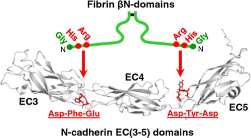
We recently identified N-cadherin as a novel receptor for fibrin and localized complementary binding sites within the fibrin βN-domains and the third and fifth extracellular domains (EC3 and EC5) of N-cadherin. We also hypothesized that the His16 and Arg17 residues of the βN-domains and the (Asp/Glu)-X-(Asp/Glu) motifs present in the EC3 and EC5 domains may play roles in the interaction between fibrin and N-cadherin. The primary objectives of this study were to test these hypotheses and to further clarify the structural basis for this interaction. To test our hypotheses, we first mutated His16 and Arg17 in the recombinant (β15–66)2 fragment, which mimics the dimeric arrangement of the βN-domains in fibrin, using site-directed mutagenesis. The results revealed that the mutations of both His16 and Arg17 are critical for the interaction. Next, we mutated Asp/Glu residues in the three (Asp/Glu)-X-(Asp/Glu) motifs, M1 (Asp-Phe-Glu), M2 (Glu-Ala-Glu), and M3 (Asp-Tyr-Asp), of the fibrin-binding N-cad(3–5) fragment of N-cadherin. The results showed that Asp292 and Glu294 of M1, and Asp468 and Asp470 of M3, are critical for the interaction. Our molecular modeling of the 3D structure of the EC3-EC4-EC5 domains revealed that these residues are located at the interfaces of EC3-EC4 and EC4-EC5 and that some may also be involved in calcium binding. In conclusion, our study identified amino acid residues in the fibrin βN-domains and the EC3 and EC5 domains of N-cadherin that are critical for the interaction of fibrin with N-cadherin and localized the fibrin-binding residues in the 3D structure of N-cadherin.
期刊介绍:
Biochemistry provides an international forum for publishing exceptional, rigorous, high-impact research across all of biological chemistry. This broad scope includes studies on the chemical, physical, mechanistic, and/or structural basis of biological or cell function, and encompasses the fields of chemical biology, synthetic biology, disease biology, cell biology, nucleic acid biology, neuroscience, structural biology, and biophysics. In addition to traditional Research Articles, Biochemistry also publishes Communications, Viewpoints, and Perspectives, as well as From the Bench articles that report new methods of particular interest to the biological chemistry community.
 求助内容:
求助内容: 应助结果提醒方式:


