Elena Hofmann, Christian Doll, Steffen Koerdt, Cynthia Kurth, Max Heiland, Kilian Kreutzer
{"title":"三维 4K 外窥镜(Orbeye™)在头颈部整形手术中的临床应用。","authors":"Elena Hofmann, Christian Doll, Steffen Koerdt, Cynthia Kurth, Max Heiland, Kilian Kreutzer","doi":"10.1007/s10006-025-01342-6","DOIUrl":null,"url":null,"abstract":"<p><strong>Purpose: </strong>To assess the clinical utility of the 3D 4K exoscope for reconstructive head and neck surgery.</p><p><strong>Methods: </strong>This retrospective study analyzed surgical details and complications with the use of the 3D 4K exoscope for microvascular reconstruction at a high-volume Department of Oral and Maxillofacial Surgery, compared to the use of a 2D microscope. Patients with oral cancer undergoing microvascular reconstruction were categorized into two cohorts based on the intraoperative use of the 3D 4K exoscope (Orbeye™, Olympus, Tokyo, Japan) or a conventional microscope (ZEISS S8 - OPMI Vario, Carl Zeiss AG, Oberkochen, Germany; Leica M680, Leica Mikrosysteme Vertrieb GmbH, Wetzlar, Germany) during a six-month study period, respectively. Outcomes were also compared between two time periods of the exoscope use to assess the learning curve over time.</p><p><strong>Results: </strong>The exoscope was applied for microvascular anastomosis in 55 surgical cases (cohort 1), and the conventional microscope was employed in 56 cases (cohort 2). The rates of postoperative complications within 14 days following the use of the exoscope were 14.5% (N = 8), compared to 16.1% (N = 9) in cohort 2. Analysis over time demonstrated a learning curve with the exoscope, reflected in a decrease in postoperative complications within 14 days from 22.7 to 9.1%.</p><p><strong>Conclusion: </strong>The three-dimensional camera system provides excellent and reliable intraoperative visualization in reconstructive head and neck surgery. Transitioning to this new technology did not lead to an increase in intra- or postoperative complications, but the successful implementation requires some experience with the device.</p>","PeriodicalId":47251,"journal":{"name":"Oral and Maxillofacial Surgery-Heidelberg","volume":"29 1","pages":"50"},"PeriodicalIF":1.8000,"publicationDate":"2025-02-03","publicationTypes":"Journal Article","fieldsOfStudy":null,"isOpenAccess":false,"openAccessPdf":"https://www.ncbi.nlm.nih.gov/pmc/articles/PMC11790798/pdf/","citationCount":"0","resultStr":"{\"title\":\"Clinical implementation of the 3D 4K exoscope (Orbeye™) in reconstructive head and neck surgery.\",\"authors\":\"Elena Hofmann, Christian Doll, Steffen Koerdt, Cynthia Kurth, Max Heiland, Kilian Kreutzer\",\"doi\":\"10.1007/s10006-025-01342-6\",\"DOIUrl\":null,\"url\":null,\"abstract\":\"<p><strong>Purpose: </strong>To assess the clinical utility of the 3D 4K exoscope for reconstructive head and neck surgery.</p><p><strong>Methods: </strong>This retrospective study analyzed surgical details and complications with the use of the 3D 4K exoscope for microvascular reconstruction at a high-volume Department of Oral and Maxillofacial Surgery, compared to the use of a 2D microscope. Patients with oral cancer undergoing microvascular reconstruction were categorized into two cohorts based on the intraoperative use of the 3D 4K exoscope (Orbeye™, Olympus, Tokyo, Japan) or a conventional microscope (ZEISS S8 - OPMI Vario, Carl Zeiss AG, Oberkochen, Germany; Leica M680, Leica Mikrosysteme Vertrieb GmbH, Wetzlar, Germany) during a six-month study period, respectively. Outcomes were also compared between two time periods of the exoscope use to assess the learning curve over time.</p><p><strong>Results: </strong>The exoscope was applied for microvascular anastomosis in 55 surgical cases (cohort 1), and the conventional microscope was employed in 56 cases (cohort 2). The rates of postoperative complications within 14 days following the use of the exoscope were 14.5% (N = 8), compared to 16.1% (N = 9) in cohort 2. Analysis over time demonstrated a learning curve with the exoscope, reflected in a decrease in postoperative complications within 14 days from 22.7 to 9.1%.</p><p><strong>Conclusion: </strong>The three-dimensional camera system provides excellent and reliable intraoperative visualization in reconstructive head and neck surgery. Transitioning to this new technology did not lead to an increase in intra- or postoperative complications, but the successful implementation requires some experience with the device.</p>\",\"PeriodicalId\":47251,\"journal\":{\"name\":\"Oral and Maxillofacial Surgery-Heidelberg\",\"volume\":\"29 1\",\"pages\":\"50\"},\"PeriodicalIF\":1.8000,\"publicationDate\":\"2025-02-03\",\"publicationTypes\":\"Journal Article\",\"fieldsOfStudy\":null,\"isOpenAccess\":false,\"openAccessPdf\":\"https://www.ncbi.nlm.nih.gov/pmc/articles/PMC11790798/pdf/\",\"citationCount\":\"0\",\"resultStr\":null,\"platform\":\"Semanticscholar\",\"paperid\":null,\"PeriodicalName\":\"Oral and Maxillofacial Surgery-Heidelberg\",\"FirstCategoryId\":\"1085\",\"ListUrlMain\":\"https://doi.org/10.1007/s10006-025-01342-6\",\"RegionNum\":0,\"RegionCategory\":null,\"ArticlePicture\":[],\"TitleCN\":null,\"AbstractTextCN\":null,\"PMCID\":null,\"EPubDate\":\"\",\"PubModel\":\"\",\"JCR\":\"Q3\",\"JCRName\":\"DENTISTRY, ORAL SURGERY & MEDICINE\",\"Score\":null,\"Total\":0}","platform":"Semanticscholar","paperid":null,"PeriodicalName":"Oral and Maxillofacial Surgery-Heidelberg","FirstCategoryId":"1085","ListUrlMain":"https://doi.org/10.1007/s10006-025-01342-6","RegionNum":0,"RegionCategory":null,"ArticlePicture":[],"TitleCN":null,"AbstractTextCN":null,"PMCID":null,"EPubDate":"","PubModel":"","JCR":"Q3","JCRName":"DENTISTRY, ORAL SURGERY & MEDICINE","Score":null,"Total":0}
Clinical implementation of the 3D 4K exoscope (Orbeye™) in reconstructive head and neck surgery.
Purpose: To assess the clinical utility of the 3D 4K exoscope for reconstructive head and neck surgery.
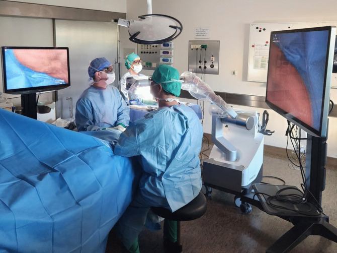
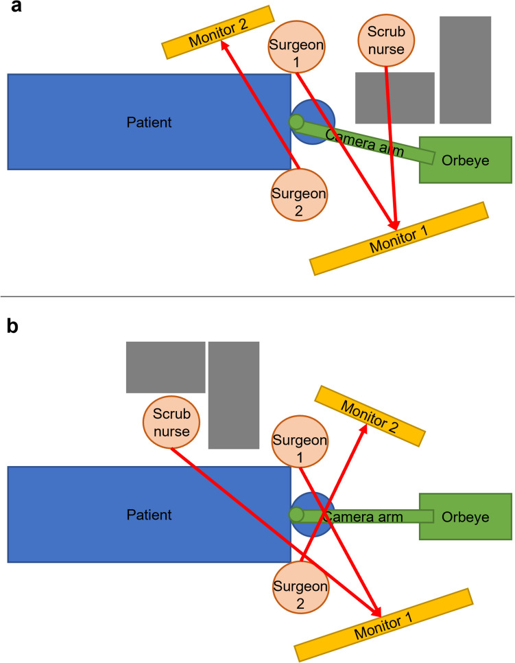
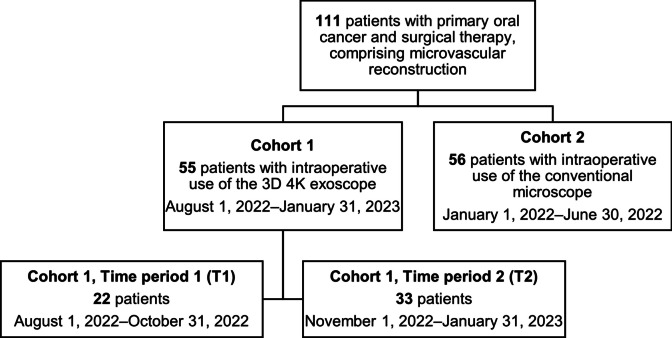
Methods: This retrospective study analyzed surgical details and complications with the use of the 3D 4K exoscope for microvascular reconstruction at a high-volume Department of Oral and Maxillofacial Surgery, compared to the use of a 2D microscope. Patients with oral cancer undergoing microvascular reconstruction were categorized into two cohorts based on the intraoperative use of the 3D 4K exoscope (Orbeye™, Olympus, Tokyo, Japan) or a conventional microscope (ZEISS S8 - OPMI Vario, Carl Zeiss AG, Oberkochen, Germany; Leica M680, Leica Mikrosysteme Vertrieb GmbH, Wetzlar, Germany) during a six-month study period, respectively. Outcomes were also compared between two time periods of the exoscope use to assess the learning curve over time.
Results: The exoscope was applied for microvascular anastomosis in 55 surgical cases (cohort 1), and the conventional microscope was employed in 56 cases (cohort 2). The rates of postoperative complications within 14 days following the use of the exoscope were 14.5% (N = 8), compared to 16.1% (N = 9) in cohort 2. Analysis over time demonstrated a learning curve with the exoscope, reflected in a decrease in postoperative complications within 14 days from 22.7 to 9.1%.
Conclusion: The three-dimensional camera system provides excellent and reliable intraoperative visualization in reconstructive head and neck surgery. Transitioning to this new technology did not lead to an increase in intra- or postoperative complications, but the successful implementation requires some experience with the device.
期刊介绍:
Oral & Maxillofacial Surgery founded as Mund-, Kiefer- und Gesichtschirurgie is a peer-reviewed online journal. It is designed for clinicians as well as researchers.The quarterly journal offers comprehensive coverage of new techniques, important developments and innovative ideas in oral and maxillofacial surgery and interdisciplinary aspects of cranial, facial and oral diseases and their management. The journal publishes papers of the highest scientific merit and widest possible scope on work in oral and maxillofacial surgery as well as supporting specialties. Practice-oriented articles help improve the methods used in oral and maxillofacial surgery.Every aspect of oral and maxillofacial surgery is fully covered through a range of invited review articles, clinical and research articles, technical notes, abstracts, and case reports. Specific topics are: aesthetic facial surgery, clinical pathology, computer-assisted surgery, congenital and craniofacial deformities, dentoalveolar surgery, head and neck oncology, implant dentistry, oral medicine, orthognathic surgery, reconstructive surgery, skull base surgery, TMJ and trauma.Time-limited reviewing and electronic processing allow to publish articles as fast as possible. Accepted articles are rapidly accessible online.Clinical studies submitted for publication have to include a declaration that they have been approved by an ethical committee according to the World Medical Association Declaration of Helsinki 1964 (last amendment during the 52nd World Medical Association General Assembly, Edinburgh, Scotland, October 2000). Experimental animal studies have to be carried out according to the principles of laboratory animal care (NIH publication No 86-23, revised 1985).
 求助内容:
求助内容: 应助结果提醒方式:


