Eyyüp Sabri Özden, Mustafa Soner Özcan, Mehtap Savran, Ilter Ilhan, Muhammet Yusuf Tepebası, Mehmet Abdulkadir Sevuk, Özlem Özmen
{"title":"塔西美酮通过NRF-2/HO-1和RIPK1/RIPK3/MLKL通路对大鼠创伤性脑损伤的影响","authors":"Eyyüp Sabri Özden, Mustafa Soner Özcan, Mehtap Savran, Ilter Ilhan, Muhammet Yusuf Tepebası, Mehmet Abdulkadir Sevuk, Özlem Özmen","doi":"10.1007/s12035-025-04711-0","DOIUrl":null,"url":null,"abstract":"<p><p>Secondary brain damageafter traumatic brain injury (TBI) involves oxidative stress, neuroinflammation, apoptosis, and necroptosis and can be reversed by understanding these molecular pathways. The objective of this study was to examine the impact of tasimelteon (Tasi) administration on brain injury through the nuclear factor erythroid 2-related factor 2 (NRF-2)/heme oxygenase-1 (HO-1) and receptor-interacting protein kinase 1 (RIPK1)/receptor-interacting protein kinase 3 (RIPK3)/mixed lineage kinase domain-like (MLKL) pathways in rats with TBI. Thirty-two male Wistar albino rats weighing 300-350 g were randomly divided into four groups: the control group, trauma group, Tasi-1 group (trauma + 1 mg/kg Tasi intraperitoneally), and Tasi-10 group (trauma + 10 mg/kg Tasi intraperitoneally). At the end of the experimental phase, after sacrifice, blood samples and brain tissue were collected for biochemical, histopathological, immunohistochemical, and genetic analyses. Tasi increased the total antioxidant status and decreased the total oxidant status and oxidative stress index. In addition, Tasi caused histopathological changes characterized by a markedly reduced hemorrhage area in the Tasi-1 group. Normal brain and meningeal structure was observed in rats in the Tasi-10 group. Immunohistochemical analysis indicated that Tasi also decreased the expression of interferon-gamma, caspase-3, and tumor necrosis factor-alpha in the brain tissue. Although NRF-2 and HO-1 expression decreased, RIPK1/RIPK3/MLKL gene expression increased due to trauma. However, Tasi treatment reversed all these findings. Tasi protected against brain injury through the NRF-2/HO-1 and RIPK1/RIPK3/MLKL pathways in rats with TBI.</p>","PeriodicalId":18762,"journal":{"name":"Molecular Neurobiology","volume":" ","pages":"12383-12392"},"PeriodicalIF":4.3000,"publicationDate":"2025-10-01","publicationTypes":"Journal Article","fieldsOfStudy":null,"isOpenAccess":false,"openAccessPdf":"https://www.ncbi.nlm.nih.gov/pmc/articles/PMC12433330/pdf/","citationCount":"0","resultStr":"{\"title\":\"Effects of Tasimelteon Treatment on Traumatic Brain Injury Through NRF-2/HO-1 and RIPK1/RIPK3/MLKL Pathways in Rats.\",\"authors\":\"Eyyüp Sabri Özden, Mustafa Soner Özcan, Mehtap Savran, Ilter Ilhan, Muhammet Yusuf Tepebası, Mehmet Abdulkadir Sevuk, Özlem Özmen\",\"doi\":\"10.1007/s12035-025-04711-0\",\"DOIUrl\":null,\"url\":null,\"abstract\":\"<p><p>Secondary brain damageafter traumatic brain injury (TBI) involves oxidative stress, neuroinflammation, apoptosis, and necroptosis and can be reversed by understanding these molecular pathways. The objective of this study was to examine the impact of tasimelteon (Tasi) administration on brain injury through the nuclear factor erythroid 2-related factor 2 (NRF-2)/heme oxygenase-1 (HO-1) and receptor-interacting protein kinase 1 (RIPK1)/receptor-interacting protein kinase 3 (RIPK3)/mixed lineage kinase domain-like (MLKL) pathways in rats with TBI. Thirty-two male Wistar albino rats weighing 300-350 g were randomly divided into four groups: the control group, trauma group, Tasi-1 group (trauma + 1 mg/kg Tasi intraperitoneally), and Tasi-10 group (trauma + 10 mg/kg Tasi intraperitoneally). At the end of the experimental phase, after sacrifice, blood samples and brain tissue were collected for biochemical, histopathological, immunohistochemical, and genetic analyses. Tasi increased the total antioxidant status and decreased the total oxidant status and oxidative stress index. In addition, Tasi caused histopathological changes characterized by a markedly reduced hemorrhage area in the Tasi-1 group. Normal brain and meningeal structure was observed in rats in the Tasi-10 group. Immunohistochemical analysis indicated that Tasi also decreased the expression of interferon-gamma, caspase-3, and tumor necrosis factor-alpha in the brain tissue. Although NRF-2 and HO-1 expression decreased, RIPK1/RIPK3/MLKL gene expression increased due to trauma. However, Tasi treatment reversed all these findings. Tasi protected against brain injury through the NRF-2/HO-1 and RIPK1/RIPK3/MLKL pathways in rats with TBI.</p>\",\"PeriodicalId\":18762,\"journal\":{\"name\":\"Molecular Neurobiology\",\"volume\":\" \",\"pages\":\"12383-12392\"},\"PeriodicalIF\":4.3000,\"publicationDate\":\"2025-10-01\",\"publicationTypes\":\"Journal Article\",\"fieldsOfStudy\":null,\"isOpenAccess\":false,\"openAccessPdf\":\"https://www.ncbi.nlm.nih.gov/pmc/articles/PMC12433330/pdf/\",\"citationCount\":\"0\",\"resultStr\":null,\"platform\":\"Semanticscholar\",\"paperid\":null,\"PeriodicalName\":\"Molecular Neurobiology\",\"FirstCategoryId\":\"3\",\"ListUrlMain\":\"https://doi.org/10.1007/s12035-025-04711-0\",\"RegionNum\":2,\"RegionCategory\":\"医学\",\"ArticlePicture\":[],\"TitleCN\":null,\"AbstractTextCN\":null,\"PMCID\":null,\"EPubDate\":\"2025/1/29 0:00:00\",\"PubModel\":\"Epub\",\"JCR\":\"Q1\",\"JCRName\":\"NEUROSCIENCES\",\"Score\":null,\"Total\":0}","platform":"Semanticscholar","paperid":null,"PeriodicalName":"Molecular Neurobiology","FirstCategoryId":"3","ListUrlMain":"https://doi.org/10.1007/s12035-025-04711-0","RegionNum":2,"RegionCategory":"医学","ArticlePicture":[],"TitleCN":null,"AbstractTextCN":null,"PMCID":null,"EPubDate":"2025/1/29 0:00:00","PubModel":"Epub","JCR":"Q1","JCRName":"NEUROSCIENCES","Score":null,"Total":0}
引用次数: 0
摘要
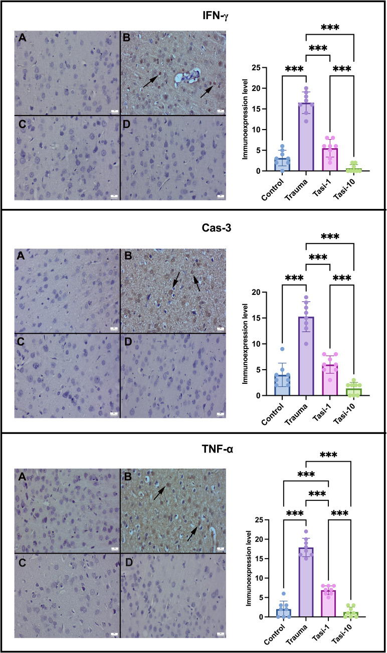
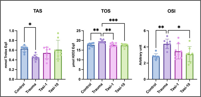
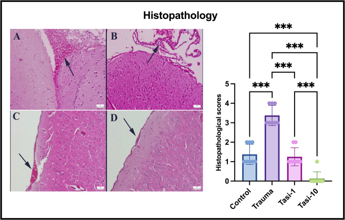
创伤性脑损伤(TBI)后的继发性脑损伤包括氧化应激、神经炎症、细胞凋亡和坏死下垂,通过了解这些分子途径可以逆转这些损伤。本研究旨在通过核因子-红细胞2相关因子2 (NRF-2)/血红素加氧酶-1 (HO-1)和受体相互作用蛋白激酶1 (RIPK1)/受体相互作用蛋白激酶3 (RIPK3)/混合谱系激酶结构域样(MLKL)通路,探讨塔西美龙(Tasi)给药对脑损伤大鼠脑损伤的影响。选取体重300 ~ 350 g的雄性Wistar白化大鼠32只,随机分为4组:对照组、创伤组、Tasi-1组(创伤+ Tasi 1 mg/kg腹腔注射)、Tasi-10组(创伤+ Tasi 10 mg/kg腹腔注射)。实验阶段结束后,取血、脑组织进行生化、组织病理学、免疫组化、遗传分析。Tasi增加了总抗氧化状态,降低了总氧化状态和氧化应激指数。此外,Tasi-1组引起的组织病理学改变以出血面积明显减少为特征。Tasi-10组大鼠脑及脑膜结构正常。免疫组织化学分析显示,Tasi还降低了脑组织中干扰素- γ、caspase-3和肿瘤坏死因子- α的表达。虽然NRF-2和HO-1表达降低,但RIPK1/RIPK3/MLKL基因表达升高。然而,Tasi治疗逆转了所有这些发现。Tasi通过NRF-2/HO-1和RIPK1/RIPK3/MLKL通路保护TBI大鼠免受脑损伤。
Effects of Tasimelteon Treatment on Traumatic Brain Injury Through NRF-2/HO-1 and RIPK1/RIPK3/MLKL Pathways in Rats.
Secondary brain damageafter traumatic brain injury (TBI) involves oxidative stress, neuroinflammation, apoptosis, and necroptosis and can be reversed by understanding these molecular pathways. The objective of this study was to examine the impact of tasimelteon (Tasi) administration on brain injury through the nuclear factor erythroid 2-related factor 2 (NRF-2)/heme oxygenase-1 (HO-1) and receptor-interacting protein kinase 1 (RIPK1)/receptor-interacting protein kinase 3 (RIPK3)/mixed lineage kinase domain-like (MLKL) pathways in rats with TBI. Thirty-two male Wistar albino rats weighing 300-350 g were randomly divided into four groups: the control group, trauma group, Tasi-1 group (trauma + 1 mg/kg Tasi intraperitoneally), and Tasi-10 group (trauma + 10 mg/kg Tasi intraperitoneally). At the end of the experimental phase, after sacrifice, blood samples and brain tissue were collected for biochemical, histopathological, immunohistochemical, and genetic analyses. Tasi increased the total antioxidant status and decreased the total oxidant status and oxidative stress index. In addition, Tasi caused histopathological changes characterized by a markedly reduced hemorrhage area in the Tasi-1 group. Normal brain and meningeal structure was observed in rats in the Tasi-10 group. Immunohistochemical analysis indicated that Tasi also decreased the expression of interferon-gamma, caspase-3, and tumor necrosis factor-alpha in the brain tissue. Although NRF-2 and HO-1 expression decreased, RIPK1/RIPK3/MLKL gene expression increased due to trauma. However, Tasi treatment reversed all these findings. Tasi protected against brain injury through the NRF-2/HO-1 and RIPK1/RIPK3/MLKL pathways in rats with TBI.
期刊介绍:
Molecular Neurobiology is an exciting journal for neuroscientists needing to stay in close touch with progress at the forefront of molecular brain research today. It is an especially important periodical for graduate students and "postdocs," specifically designed to synthesize and critically assess research trends for all neuroscientists hoping to stay active at the cutting edge of this dramatically developing area. This journal has proven to be crucial in departmental libraries, serving as essential reading for every committed neuroscientist who is striving to keep abreast of all rapid developments in a forefront field. Most recent significant advances in experimental and clinical neuroscience have been occurring at the molecular level. Until now, there has been no journal devoted to looking closely at this fragmented literature in a critical, coherent fashion. Each submission is thoroughly analyzed by scientists and clinicians internationally renowned for their special competence in the areas treated.
 求助内容:
求助内容: 应助结果提醒方式:


