{"title":"肱-踝脉波速度反映腹主动脉瘤患者局部动脉僵硬性和扩张性。","authors":"Toshiya Nishibe, Shinobu Akiyama, Masaki Kano, Shoji Fukuda, Fumio Chiba, Jun Koizumi, Masayasu Nishibe","doi":"10.3400/avd.oa.24-00097","DOIUrl":null,"url":null,"abstract":"<p><p><b>Objectives:</b> We investigated the association between brachial-ankle pulse wave velocity (PWV) and arterial stiffness and distensibility in the aneurysmal sac of abdominal aortic aneurysm (AAA). <b>Methods:</b> Data from 49 patients with AAA from June 2020 to November 2022 at Tokyo Medical University Hospital were retrospectively analyzed. Brachial-ankle PWV (cm/s) was obtained via an automated oscillometric method. Regional arterial stiffness and distensibility parameters, such as stiffness parameter (β), pressure-strain elasticity modulus (Ep, kPa), one-point PWV (PWV β, m/s), and arterial compliance (AC, mm<sup>2</sup>/kPa<sup>-1</sup>), were assessed using 2-dimensional automated tissue tracking (2DTT) ultrasonography. Patients were divided into two groups: high PWV (≥1800) and low PWV (<1800). <b>Results:</b> Patients with high PWV showed significantly higher β and PWV β (30.6 ± 10.1 vs. 25.2 ± 6.3, p = 0.047; 11.6 ± 2.3 vs. 10.5 ± 1.5, p = 0.048) and significantly lower AC in the aneurysmal sac (10.6 ± 5.3 vs. 14.7 ± 8.1, p = 0.045) than those with low PWV. AC was negatively correlated with PWV (r = -0.361, p = 0.011). <b>Conclusions:</b> Brachial-ankle PWV can reflect arterial stiffness and distensibility, as measured by 2DTT ultrasonography, in the aneurysmal sac of AAA, suggesting its potential as an elasticity index for assessing regional arterial stiffness and distensibility in AAA.</p>","PeriodicalId":7995,"journal":{"name":"Annals of vascular diseases","volume":"18 1","pages":""},"PeriodicalIF":0.6000,"publicationDate":"2025-01-01","publicationTypes":"Journal Article","fieldsOfStudy":null,"isOpenAccess":false,"openAccessPdf":"https://www.ncbi.nlm.nih.gov/pmc/articles/PMC11771152/pdf/","citationCount":"0","resultStr":"{\"title\":\"Brachial-Ankle Pulse Wave Velocity Reflects Regional Arterial Stiffness and Distensibility in Patients with Abdominal Aortic Aneurysm.\",\"authors\":\"Toshiya Nishibe, Shinobu Akiyama, Masaki Kano, Shoji Fukuda, Fumio Chiba, Jun Koizumi, Masayasu Nishibe\",\"doi\":\"10.3400/avd.oa.24-00097\",\"DOIUrl\":null,\"url\":null,\"abstract\":\"<p><p><b>Objectives:</b> We investigated the association between brachial-ankle pulse wave velocity (PWV) and arterial stiffness and distensibility in the aneurysmal sac of abdominal aortic aneurysm (AAA). <b>Methods:</b> Data from 49 patients with AAA from June 2020 to November 2022 at Tokyo Medical University Hospital were retrospectively analyzed. Brachial-ankle PWV (cm/s) was obtained via an automated oscillometric method. Regional arterial stiffness and distensibility parameters, such as stiffness parameter (β), pressure-strain elasticity modulus (Ep, kPa), one-point PWV (PWV β, m/s), and arterial compliance (AC, mm<sup>2</sup>/kPa<sup>-1</sup>), were assessed using 2-dimensional automated tissue tracking (2DTT) ultrasonography. Patients were divided into two groups: high PWV (≥1800) and low PWV (<1800). <b>Results:</b> Patients with high PWV showed significantly higher β and PWV β (30.6 ± 10.1 vs. 25.2 ± 6.3, p = 0.047; 11.6 ± 2.3 vs. 10.5 ± 1.5, p = 0.048) and significantly lower AC in the aneurysmal sac (10.6 ± 5.3 vs. 14.7 ± 8.1, p = 0.045) than those with low PWV. AC was negatively correlated with PWV (r = -0.361, p = 0.011). <b>Conclusions:</b> Brachial-ankle PWV can reflect arterial stiffness and distensibility, as measured by 2DTT ultrasonography, in the aneurysmal sac of AAA, suggesting its potential as an elasticity index for assessing regional arterial stiffness and distensibility in AAA.</p>\",\"PeriodicalId\":7995,\"journal\":{\"name\":\"Annals of vascular diseases\",\"volume\":\"18 1\",\"pages\":\"\"},\"PeriodicalIF\":0.6000,\"publicationDate\":\"2025-01-01\",\"publicationTypes\":\"Journal Article\",\"fieldsOfStudy\":null,\"isOpenAccess\":false,\"openAccessPdf\":\"https://www.ncbi.nlm.nih.gov/pmc/articles/PMC11771152/pdf/\",\"citationCount\":\"0\",\"resultStr\":null,\"platform\":\"Semanticscholar\",\"paperid\":null,\"PeriodicalName\":\"Annals of vascular diseases\",\"FirstCategoryId\":\"1085\",\"ListUrlMain\":\"https://doi.org/10.3400/avd.oa.24-00097\",\"RegionNum\":0,\"RegionCategory\":null,\"ArticlePicture\":[],\"TitleCN\":null,\"AbstractTextCN\":null,\"PMCID\":null,\"EPubDate\":\"\",\"PubModel\":\"\",\"JCR\":\"Q4\",\"JCRName\":\"PERIPHERAL VASCULAR DISEASE\",\"Score\":null,\"Total\":0}","platform":"Semanticscholar","paperid":null,"PeriodicalName":"Annals of vascular diseases","FirstCategoryId":"1085","ListUrlMain":"https://doi.org/10.3400/avd.oa.24-00097","RegionNum":0,"RegionCategory":null,"ArticlePicture":[],"TitleCN":null,"AbstractTextCN":null,"PMCID":null,"EPubDate":"","PubModel":"","JCR":"Q4","JCRName":"PERIPHERAL VASCULAR DISEASE","Score":null,"Total":0}
引用次数: 0
摘要
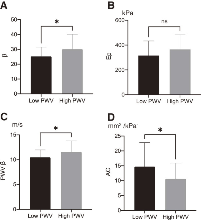
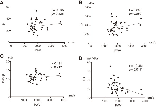
目的:探讨腹主动脉瘤(AAA)动脉瘤囊内脉波速度(PWV)与动脉僵硬度和扩张度的关系。方法:回顾性分析2020年6月至2022年11月东京医科大学医院49例AAA患者的资料。通过自动示波法获得肱-踝关节PWV (cm/s)。采用二维自动组织跟踪(2DTT)超声评估区域动脉刚度和扩张参数,如刚度参数(β)、压力-应变弹性模量(Ep, kPa)、一点PWV (PWV β, m/s)和动脉顺应性(AC, mm2/kPa-1)。患者分为高PWV组(≥1800)和低PWV组(结果:高PWV组患者β和PWV β显著升高(30.6±10.1∶25.2±6.3,p = 0.047;(11.6±2.3 vs. 10.5±1.5,p = 0.048),与低PWV组相比,动脉瘤囊AC明显降低(10.6±5.3 vs. 14.7±8.1,p = 0.045)。AC与PWV呈负相关(r = -0.361, p = 0.011)。结论:2DTT超声测量肱踝PWV能反映AAA动脉瘤囊内动脉僵硬度和扩张度,提示其可作为评估AAA局部动脉僵硬度和扩张度的弹性指标。
Brachial-Ankle Pulse Wave Velocity Reflects Regional Arterial Stiffness and Distensibility in Patients with Abdominal Aortic Aneurysm.
Objectives: We investigated the association between brachial-ankle pulse wave velocity (PWV) and arterial stiffness and distensibility in the aneurysmal sac of abdominal aortic aneurysm (AAA). Methods: Data from 49 patients with AAA from June 2020 to November 2022 at Tokyo Medical University Hospital were retrospectively analyzed. Brachial-ankle PWV (cm/s) was obtained via an automated oscillometric method. Regional arterial stiffness and distensibility parameters, such as stiffness parameter (β), pressure-strain elasticity modulus (Ep, kPa), one-point PWV (PWV β, m/s), and arterial compliance (AC, mm2/kPa-1), were assessed using 2-dimensional automated tissue tracking (2DTT) ultrasonography. Patients were divided into two groups: high PWV (≥1800) and low PWV (<1800). Results: Patients with high PWV showed significantly higher β and PWV β (30.6 ± 10.1 vs. 25.2 ± 6.3, p = 0.047; 11.6 ± 2.3 vs. 10.5 ± 1.5, p = 0.048) and significantly lower AC in the aneurysmal sac (10.6 ± 5.3 vs. 14.7 ± 8.1, p = 0.045) than those with low PWV. AC was negatively correlated with PWV (r = -0.361, p = 0.011). Conclusions: Brachial-ankle PWV can reflect arterial stiffness and distensibility, as measured by 2DTT ultrasonography, in the aneurysmal sac of AAA, suggesting its potential as an elasticity index for assessing regional arterial stiffness and distensibility in AAA.

 求助内容:
求助内容: 应助结果提醒方式:


