Nathan P Coles, Suzan Elsheikh, Agathe Quesnel, Lucy Butler, Claire Jennings, Chaimaa Tarzi, Ojodomo J Achadu, Meez Islam, Karunakaran Kalesh, Annalisa Occhipinti, Claudio Angione, Jon Marles-Wright, David J Koss, Alan J Thomas, Tiago F Outeiro, Panagiota S Filippou, Ahmad A Khundakar
{"title":"分子洞察α-突触核蛋白颤动:拉曼光谱和机器学习方法。","authors":"Nathan P Coles, Suzan Elsheikh, Agathe Quesnel, Lucy Butler, Claire Jennings, Chaimaa Tarzi, Ojodomo J Achadu, Meez Islam, Karunakaran Kalesh, Annalisa Occhipinti, Claudio Angione, Jon Marles-Wright, David J Koss, Alan J Thomas, Tiago F Outeiro, Panagiota S Filippou, Ahmad A Khundakar","doi":"10.1021/acschemneuro.4c00726","DOIUrl":null,"url":null,"abstract":"<p><p>The aggregation of α-synuclein is crucial to the development of Lewy body diseases, including Parkinson's disease and dementia with Lewy bodies. The aggregation pathway of α-synuclein typically involves a defined sequence of nucleation, elongation, and secondary nucleation, exhibiting prion-like spreading. This study employed Raman spectroscopy and machine learning analysis, alongside complementary techniques, to characterize the biomolecular changes during the fibrillation of purified recombinant wild-type α-synuclein protein. Monomeric α-synuclein was produced, purified, and subjected to a 7-day fibrillation assay to generate preformed fibrils. Stages of α-synuclein fibrillation were analyzed using Raman spectroscopy, with aggregation confirmed through negative staining transmission electron microscopy, mass spectrometry, and light scattering analyses. A machine learning pipeline incorporating principal component analysis and uniform manifold approximation and projection was used to analyze the Raman spectral data and identify significant peaks, resulting in differentiation between sample groups. Notable spectral shifts in α-synuclein were found in various stages of aggregation. Early changes (D1) included increases in α-helical structures (1303, 1330 cm<sup>-1</sup>) and β-sheet formation (1045 cm<sup>-1</sup>), with reductions in COO<sup>-</sup> and CH<sub>2</sub> bond regions (1406, 1445 cm<sup>-1</sup>). By D4, these structural shifts persist with additional β-sheet features. At D7, a decrease in β-sheet H-bonding (1625 cm<sup>-1</sup>) and tyrosine ring breathing (830 cm<sup>-1</sup>) indicates further structural stabilization, suggesting a shift from initial helical structures to stabilized β-sheets and aggregated fibrils. Additionally, alterations in peaks related to tyrosine, alanine, proline, and glutamic acid were identified, emphasizing the role of these amino acids in intramolecular interactions during the transition from α-helical to β-sheet conformational states in α-synuclein fibrillation. This approach offers insight into α-synuclein aggregation, enhancing the understanding of its role in Lewy body disease pathophysiology and potential diagnostic relevance.</p>","PeriodicalId":13,"journal":{"name":"ACS Chemical Neuroscience","volume":" ","pages":"687-698"},"PeriodicalIF":3.9000,"publicationDate":"2025-02-19","publicationTypes":"Journal Article","fieldsOfStudy":null,"isOpenAccess":false,"openAccessPdf":"https://www.ncbi.nlm.nih.gov/pmc/articles/PMC11843597/pdf/","citationCount":"0","resultStr":"{\"title\":\"Molecular Insights into α-Synuclein Fibrillation: A Raman Spectroscopy and Machine Learning Approach.\",\"authors\":\"Nathan P Coles, Suzan Elsheikh, Agathe Quesnel, Lucy Butler, Claire Jennings, Chaimaa Tarzi, Ojodomo J Achadu, Meez Islam, Karunakaran Kalesh, Annalisa Occhipinti, Claudio Angione, Jon Marles-Wright, David J Koss, Alan J Thomas, Tiago F Outeiro, Panagiota S Filippou, Ahmad A Khundakar\",\"doi\":\"10.1021/acschemneuro.4c00726\",\"DOIUrl\":null,\"url\":null,\"abstract\":\"<p><p>The aggregation of α-synuclein is crucial to the development of Lewy body diseases, including Parkinson's disease and dementia with Lewy bodies. The aggregation pathway of α-synuclein typically involves a defined sequence of nucleation, elongation, and secondary nucleation, exhibiting prion-like spreading. This study employed Raman spectroscopy and machine learning analysis, alongside complementary techniques, to characterize the biomolecular changes during the fibrillation of purified recombinant wild-type α-synuclein protein. Monomeric α-synuclein was produced, purified, and subjected to a 7-day fibrillation assay to generate preformed fibrils. Stages of α-synuclein fibrillation were analyzed using Raman spectroscopy, with aggregation confirmed through negative staining transmission electron microscopy, mass spectrometry, and light scattering analyses. A machine learning pipeline incorporating principal component analysis and uniform manifold approximation and projection was used to analyze the Raman spectral data and identify significant peaks, resulting in differentiation between sample groups. Notable spectral shifts in α-synuclein were found in various stages of aggregation. Early changes (D1) included increases in α-helical structures (1303, 1330 cm<sup>-1</sup>) and β-sheet formation (1045 cm<sup>-1</sup>), with reductions in COO<sup>-</sup> and CH<sub>2</sub> bond regions (1406, 1445 cm<sup>-1</sup>). By D4, these structural shifts persist with additional β-sheet features. At D7, a decrease in β-sheet H-bonding (1625 cm<sup>-1</sup>) and tyrosine ring breathing (830 cm<sup>-1</sup>) indicates further structural stabilization, suggesting a shift from initial helical structures to stabilized β-sheets and aggregated fibrils. Additionally, alterations in peaks related to tyrosine, alanine, proline, and glutamic acid were identified, emphasizing the role of these amino acids in intramolecular interactions during the transition from α-helical to β-sheet conformational states in α-synuclein fibrillation. This approach offers insight into α-synuclein aggregation, enhancing the understanding of its role in Lewy body disease pathophysiology and potential diagnostic relevance.</p>\",\"PeriodicalId\":13,\"journal\":{\"name\":\"ACS Chemical Neuroscience\",\"volume\":\" \",\"pages\":\"687-698\"},\"PeriodicalIF\":3.9000,\"publicationDate\":\"2025-02-19\",\"publicationTypes\":\"Journal Article\",\"fieldsOfStudy\":null,\"isOpenAccess\":false,\"openAccessPdf\":\"https://www.ncbi.nlm.nih.gov/pmc/articles/PMC11843597/pdf/\",\"citationCount\":\"0\",\"resultStr\":null,\"platform\":\"Semanticscholar\",\"paperid\":null,\"PeriodicalName\":\"ACS Chemical Neuroscience\",\"FirstCategoryId\":\"3\",\"ListUrlMain\":\"https://doi.org/10.1021/acschemneuro.4c00726\",\"RegionNum\":3,\"RegionCategory\":\"医学\",\"ArticlePicture\":[],\"TitleCN\":null,\"AbstractTextCN\":null,\"PMCID\":null,\"EPubDate\":\"2025/1/28 0:00:00\",\"PubModel\":\"Epub\",\"JCR\":\"Q2\",\"JCRName\":\"BIOCHEMISTRY & MOLECULAR BIOLOGY\",\"Score\":null,\"Total\":0}","platform":"Semanticscholar","paperid":null,"PeriodicalName":"ACS Chemical Neuroscience","FirstCategoryId":"3","ListUrlMain":"https://doi.org/10.1021/acschemneuro.4c00726","RegionNum":3,"RegionCategory":"医学","ArticlePicture":[],"TitleCN":null,"AbstractTextCN":null,"PMCID":null,"EPubDate":"2025/1/28 0:00:00","PubModel":"Epub","JCR":"Q2","JCRName":"BIOCHEMISTRY & MOLECULAR BIOLOGY","Score":null,"Total":0}
Molecular Insights into α-Synuclein Fibrillation: A Raman Spectroscopy and Machine Learning Approach.
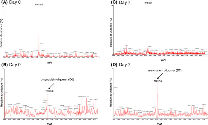
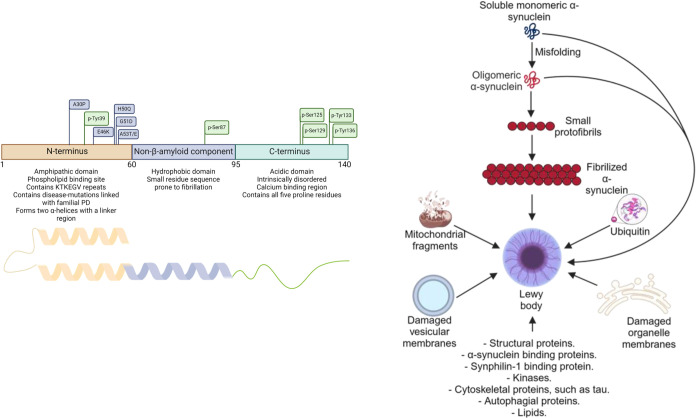
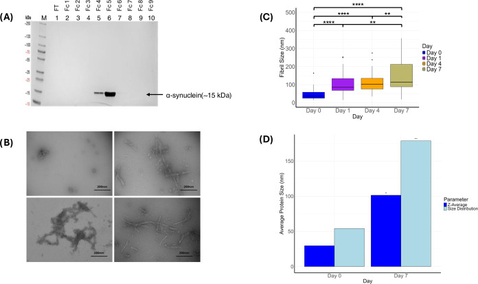
The aggregation of α-synuclein is crucial to the development of Lewy body diseases, including Parkinson's disease and dementia with Lewy bodies. The aggregation pathway of α-synuclein typically involves a defined sequence of nucleation, elongation, and secondary nucleation, exhibiting prion-like spreading. This study employed Raman spectroscopy and machine learning analysis, alongside complementary techniques, to characterize the biomolecular changes during the fibrillation of purified recombinant wild-type α-synuclein protein. Monomeric α-synuclein was produced, purified, and subjected to a 7-day fibrillation assay to generate preformed fibrils. Stages of α-synuclein fibrillation were analyzed using Raman spectroscopy, with aggregation confirmed through negative staining transmission electron microscopy, mass spectrometry, and light scattering analyses. A machine learning pipeline incorporating principal component analysis and uniform manifold approximation and projection was used to analyze the Raman spectral data and identify significant peaks, resulting in differentiation between sample groups. Notable spectral shifts in α-synuclein were found in various stages of aggregation. Early changes (D1) included increases in α-helical structures (1303, 1330 cm-1) and β-sheet formation (1045 cm-1), with reductions in COO- and CH2 bond regions (1406, 1445 cm-1). By D4, these structural shifts persist with additional β-sheet features. At D7, a decrease in β-sheet H-bonding (1625 cm-1) and tyrosine ring breathing (830 cm-1) indicates further structural stabilization, suggesting a shift from initial helical structures to stabilized β-sheets and aggregated fibrils. Additionally, alterations in peaks related to tyrosine, alanine, proline, and glutamic acid were identified, emphasizing the role of these amino acids in intramolecular interactions during the transition from α-helical to β-sheet conformational states in α-synuclein fibrillation. This approach offers insight into α-synuclein aggregation, enhancing the understanding of its role in Lewy body disease pathophysiology and potential diagnostic relevance.
期刊介绍:
ACS Chemical Neuroscience publishes high-quality research articles and reviews that showcase chemical, quantitative biological, biophysical and bioengineering approaches to the understanding of the nervous system and to the development of new treatments for neurological disorders. Research in the journal focuses on aspects of chemical neurobiology and bio-neurochemistry such as the following:
Neurotransmitters and receptors
Neuropharmaceuticals and therapeutics
Neural development—Plasticity, and degeneration
Chemical, physical, and computational methods in neuroscience
Neuronal diseases—basis, detection, and treatment
Mechanism of aging, learning, memory and behavior
Pain and sensory processing
Neurotoxins
Neuroscience-inspired bioengineering
Development of methods in chemical neurobiology
Neuroimaging agents and technologies
Animal models for central nervous system diseases
Behavioral research
 求助内容:
求助内容: 应助结果提醒方式:


